Yi Hong Li, Zhon Min Huang, Ji Kuen Yu, Yi Sheng Lin, Chao Yu Hsu, Min Che Tung
{"title":"Misdiagnosis of vasitis: a potential diagnostic pitfall with computed tomography.","authors":"Yi Hong Li, Zhon Min Huang, Ji Kuen Yu, Yi Sheng Lin, Chao Yu Hsu, Min Che Tung","doi":"10.1186/s12610-022-00168-6","DOIUrl":null,"url":null,"abstract":"<p><strong>Background: </strong>Vasitis is a rare condition that may be challenging for the clinical practitioner. Sometimes it is misdiagnosed as incarcerated inguinal hernia; thus, patients end up receiving unnecessary surgery. Compared with the traditional approach with only sonography, the more recent introduction of computed tomography in the diagnostic process has provided higher quality imaging and more detailed anatomy. Consequently, some urologists advocate the efficacy of computed tomography in the differential diagnosis of difficult cases.</p><p><strong>Case presentation: </strong>We present the case of a 23-year-old male who suffered from right inguinal pain and swelling. His scrotum ultrasound showed multiple tubular structure dilatation within the subinguinal area and no testis torsion. The initial diagnosis was a right inguinal hernia. Computed tomography supported that initial diagnosis, and we presumed the lesion represented a herniation of the omentum with mesenteric vessels. Since there was a suspicion of hernia incarceration, the patient underwent diagnostic laparoscopy, which did not reveal herniation, but only erythematous reaction and swelling over the right spermatic cord. Following a final diagnosis of vasitis, he received empirical antibiotic treatment and his symptoms entirely resolved.</p><p><strong>Conclusions: </strong>Even though computed tomography can provide thorough imaging of the urogenital system, the contrast enhancement within vessels and inflammatory organs can still be misleading in the diagnostic process.</p>","PeriodicalId":8730,"journal":{"name":"Basic and Clinical Andrology","volume":" ","pages":"19"},"PeriodicalIF":2.4000,"publicationDate":"2022-10-11","publicationTypes":"Journal Article","fieldsOfStudy":null,"isOpenAccess":false,"openAccessPdf":"https://www.ncbi.nlm.nih.gov/pmc/articles/PMC9552416/pdf/","citationCount":"0","resultStr":null,"platform":"Semanticscholar","paperid":null,"PeriodicalName":"Basic and Clinical Andrology","FirstCategoryId":"3","ListUrlMain":"https://doi.org/10.1186/s12610-022-00168-6","RegionNum":3,"RegionCategory":"医学","ArticlePicture":[],"TitleCN":null,"AbstractTextCN":null,"PMCID":null,"EPubDate":"","PubModel":"","JCR":"Q2","JCRName":"ANDROLOGY","Score":null,"Total":0}
引用次数: 0
Abstract
Background: Vasitis is a rare condition that may be challenging for the clinical practitioner. Sometimes it is misdiagnosed as incarcerated inguinal hernia; thus, patients end up receiving unnecessary surgery. Compared with the traditional approach with only sonography, the more recent introduction of computed tomography in the diagnostic process has provided higher quality imaging and more detailed anatomy. Consequently, some urologists advocate the efficacy of computed tomography in the differential diagnosis of difficult cases.
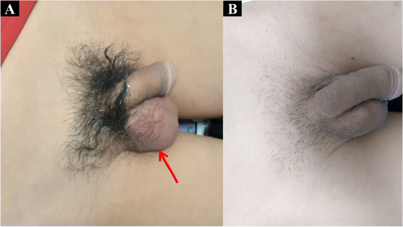
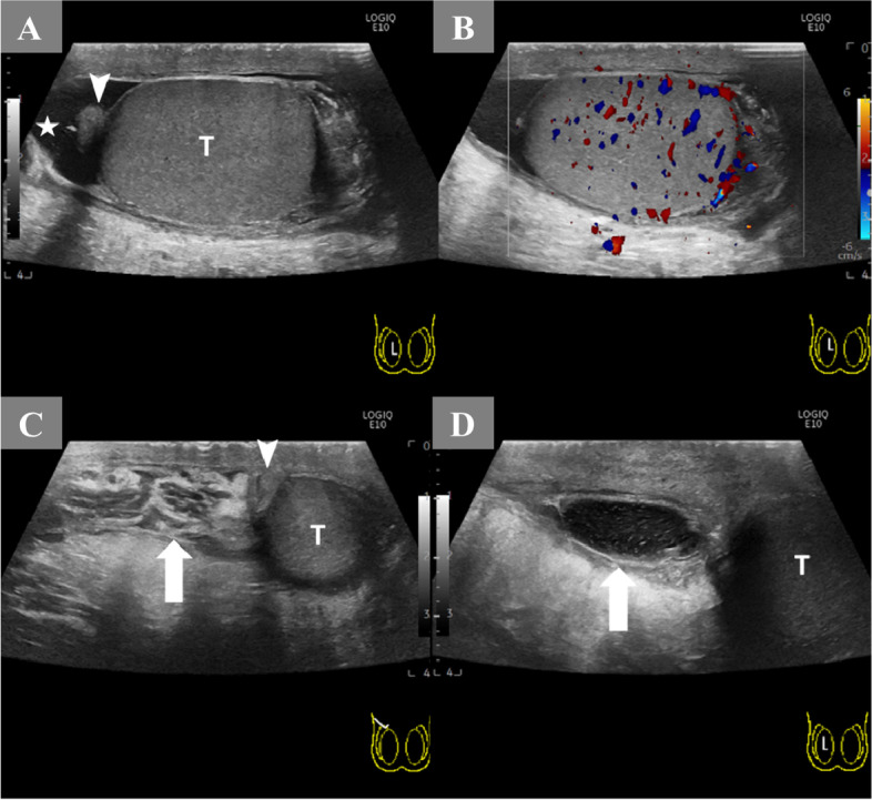
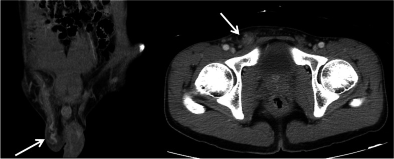
Case presentation: We present the case of a 23-year-old male who suffered from right inguinal pain and swelling. His scrotum ultrasound showed multiple tubular structure dilatation within the subinguinal area and no testis torsion. The initial diagnosis was a right inguinal hernia. Computed tomography supported that initial diagnosis, and we presumed the lesion represented a herniation of the omentum with mesenteric vessels. Since there was a suspicion of hernia incarceration, the patient underwent diagnostic laparoscopy, which did not reveal herniation, but only erythematous reaction and swelling over the right spermatic cord. Following a final diagnosis of vasitis, he received empirical antibiotic treatment and his symptoms entirely resolved.
Conclusions: Even though computed tomography can provide thorough imaging of the urogenital system, the contrast enhancement within vessels and inflammatory organs can still be misleading in the diagnostic process.
期刊介绍:
Basic and Clinical Andrology is an open access journal in the domain of andrology covering all aspects of male reproductive and sexual health in both human and animal models. The journal aims to bring to light the various clinical advancements and research developments in andrology from the international community.
 求助内容:
求助内容: 应助结果提醒方式:


