{"title":"Spotted Temporal Lobe Necrosis following Concurrent Chemoradiation Therapy Using Image-Guided Radiotherapy for Nasopharyngeal Carcinoma.","authors":"Yu-Wei Chiang, Li-Jen Liao, Chia-Yun Wu, Wu-Chia Lo, Pei-Wei Shueng, Chen-Xiong Hsu, Deng-Yu Guo, Pei-Yu Hou, Pei-Ying Hsieh, Chen-Hsi Hsieh","doi":"10.1155/2022/5877106","DOIUrl":null,"url":null,"abstract":"<p><strong>Background: </strong>To explore spotted temporal lobe necrosis (TLN) and changes in brain magnetic resonance imaging (MRI) after image-guided radiotherapy (IGRT) in a patient with nasopharyngeal carcinoma (NPC). Case presentation: a 57-year-old male was diagnosed with stage III NPC, cT1N2M0, in 2017. He underwent concurrent chemoradiation therapy (CCRT) with cisplatin (30 mg/m<sup>2</sup>) and 5- fluorouracil (5-FU, 500 mg/m<sup>2</sup>) plus IGRT with 70 Gy in 35 fractions for 7 weeks. The following MRI showed a complete response in the NPC. However, the patient suffered from fainting periodically when standing up approximately 3 years after CCRT. Neck sonography showed mild atherosclerosis (< 15%) of bilateral carotid bifurcations and bilateral small-diameter vertebral arteries, with reduced flow volume. The following MRI showed a 9 mm × 7 mm enhancing lesion in the right temporal lobe without locoregional recurrence, and TLN was diagnosed. The lesion was near the watershed area between the anterior temporal and temporo-occipital arteries. The volume of the necrotic lesion was 0.51 c.c., and the mean dose and Dmax of the lesion were 64.4 Gy and 73.7 Gy, respectively. Additionally, the mean dose, V45, D1 c.c. (dose to 1 ml of the temporal lobe volume), D0.5 c.c. and Dmax of the right and left temporal lobes were 11.1 Gy and 11.4 Gy, 8.5 c.c. and 6.7 c.c., 70.1 Gy and 67.1 Gy, 72.0 Gy and 68.8 Gy, and 74.2 Gy and 72.1 Gy, respectively.</p><p><strong>Conclusion: </strong>Spotted TLN in patients with NPC treated by IGRT may be difficult to diagnose due to a lack of clinical symptoms and radiological signs. Endothelial damage may occur in carotid and vertebral arteries within the irradiated area, affecting the small branches supplying the temporal lobe and inducing spotted TLN. Future research on the relationship between vessels and RT or CCRT and the development of TLN is warranted.</p>","PeriodicalId":45872,"journal":{"name":"Case Reports in Otolaryngology","volume":"2022 ","pages":"5877106"},"PeriodicalIF":0.4000,"publicationDate":"2022-09-27","publicationTypes":"Journal Article","fieldsOfStudy":null,"isOpenAccess":false,"openAccessPdf":"https://www.ncbi.nlm.nih.gov/pmc/articles/PMC9532156/pdf/","citationCount":"1","resultStr":null,"platform":"Semanticscholar","paperid":null,"PeriodicalName":"Case Reports in Otolaryngology","FirstCategoryId":"1085","ListUrlMain":"https://doi.org/10.1155/2022/5877106","RegionNum":0,"RegionCategory":null,"ArticlePicture":[],"TitleCN":null,"AbstractTextCN":null,"PMCID":null,"EPubDate":"2022/1/1 0:00:00","PubModel":"eCollection","JCR":"Q4","JCRName":"OTORHINOLARYNGOLOGY","Score":null,"Total":0}
引用次数: 1
Abstract
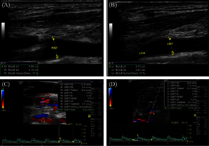
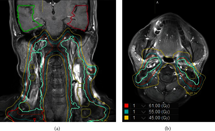
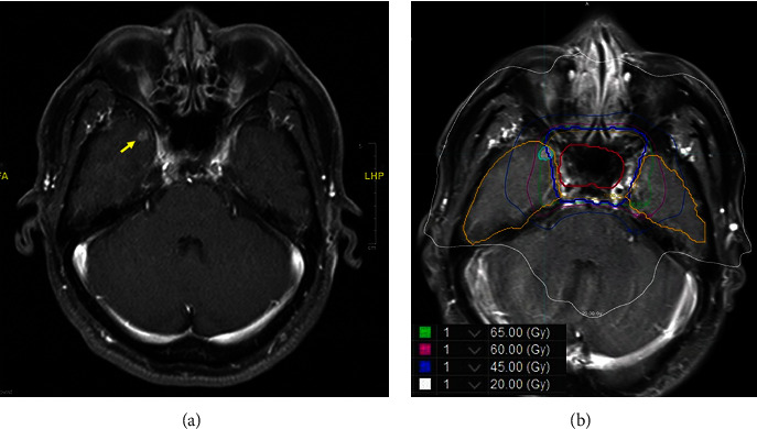
Background: To explore spotted temporal lobe necrosis (TLN) and changes in brain magnetic resonance imaging (MRI) after image-guided radiotherapy (IGRT) in a patient with nasopharyngeal carcinoma (NPC). Case presentation: a 57-year-old male was diagnosed with stage III NPC, cT1N2M0, in 2017. He underwent concurrent chemoradiation therapy (CCRT) with cisplatin (30 mg/m2) and 5- fluorouracil (5-FU, 500 mg/m2) plus IGRT with 70 Gy in 35 fractions for 7 weeks. The following MRI showed a complete response in the NPC. However, the patient suffered from fainting periodically when standing up approximately 3 years after CCRT. Neck sonography showed mild atherosclerosis (< 15%) of bilateral carotid bifurcations and bilateral small-diameter vertebral arteries, with reduced flow volume. The following MRI showed a 9 mm × 7 mm enhancing lesion in the right temporal lobe without locoregional recurrence, and TLN was diagnosed. The lesion was near the watershed area between the anterior temporal and temporo-occipital arteries. The volume of the necrotic lesion was 0.51 c.c., and the mean dose and Dmax of the lesion were 64.4 Gy and 73.7 Gy, respectively. Additionally, the mean dose, V45, D1 c.c. (dose to 1 ml of the temporal lobe volume), D0.5 c.c. and Dmax of the right and left temporal lobes were 11.1 Gy and 11.4 Gy, 8.5 c.c. and 6.7 c.c., 70.1 Gy and 67.1 Gy, 72.0 Gy and 68.8 Gy, and 74.2 Gy and 72.1 Gy, respectively.
Conclusion: Spotted TLN in patients with NPC treated by IGRT may be difficult to diagnose due to a lack of clinical symptoms and radiological signs. Endothelial damage may occur in carotid and vertebral arteries within the irradiated area, affecting the small branches supplying the temporal lobe and inducing spotted TLN. Future research on the relationship between vessels and RT or CCRT and the development of TLN is warranted.

 求助内容:
求助内容: 应助结果提醒方式:


