J Altenbernd, S Zimmer, L Andrae, B Labonte, J Gruber, H Beier, M Abdulgader, M Buechter, M Forsting, J Theysohn
{"title":"High volume retrograde portography for better discrimination of the portal vein during TIPS procedure.","authors":"J Altenbernd, S Zimmer, L Andrae, B Labonte, J Gruber, H Beier, M Abdulgader, M Buechter, M Forsting, J Theysohn","doi":"10.1177/20584601221128405","DOIUrl":null,"url":null,"abstract":"<p><p><b>Background:</b> Imaging of the portal vein prior to puncture for TIPS is essential. <b>Purpose:</b> With this study, we examined a modified retrograde portography with regard to the reliable representation of the portal vein. <b>Material and Methods:</b> Prospective evaluation of 65 TIPS interventions with regard to the delimitation of the portal vein and the exact parameters of retrograde portography such as catheter diameter and contrast medium volume per injection. <b>Results:</b> Retrograde portographies with a large-lumen catheter (10 F) and a large contrast medium volume (40 mL) were performed in 35/63 patients with significantly better delineation of the portal vein than when using 5 F catheters with 10 mL contrast medium. <b>Conclusion:</b> The so-called high volume retrograde portography leads to better delimitation of the portal vein during TIPS application.</p>","PeriodicalId":72063,"journal":{"name":"Acta radiologica open","volume":" ","pages":"20584601221128405"},"PeriodicalIF":1.0000,"publicationDate":"2022-09-20","publicationTypes":"Journal Article","fieldsOfStudy":null,"isOpenAccess":false,"openAccessPdf":"https://www.ncbi.nlm.nih.gov/pmc/articles/PMC9493682/pdf/","citationCount":"0","resultStr":null,"platform":"Semanticscholar","paperid":null,"PeriodicalName":"Acta radiologica open","FirstCategoryId":"1085","ListUrlMain":"https://doi.org/10.1177/20584601221128405","RegionNum":0,"RegionCategory":null,"ArticlePicture":[],"TitleCN":null,"AbstractTextCN":null,"PMCID":null,"EPubDate":"2022/9/1 0:00:00","PubModel":"eCollection","JCR":"Q4","JCRName":"RADIOLOGY, NUCLEAR MEDICINE & MEDICAL IMAGING","Score":null,"Total":0}
引用次数: 0
Abstract
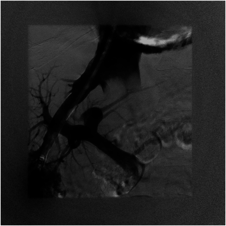
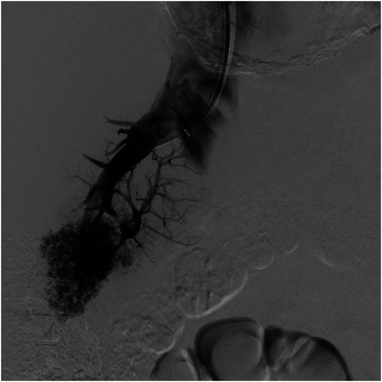
Background: Imaging of the portal vein prior to puncture for TIPS is essential. Purpose: With this study, we examined a modified retrograde portography with regard to the reliable representation of the portal vein. Material and Methods: Prospective evaluation of 65 TIPS interventions with regard to the delimitation of the portal vein and the exact parameters of retrograde portography such as catheter diameter and contrast medium volume per injection. Results: Retrograde portographies with a large-lumen catheter (10 F) and a large contrast medium volume (40 mL) were performed in 35/63 patients with significantly better delineation of the portal vein than when using 5 F catheters with 10 mL contrast medium. Conclusion: The so-called high volume retrograde portography leads to better delimitation of the portal vein during TIPS application.

 求助内容:
求助内容: 应助结果提醒方式:


