Ramesh Venkatesh, Aditi Gupta, Naresh Kumar Yadav, Jay Chhablani
{"title":"Presumed Combined Brilliant Blue G and Endolight-Induced Macular Damage following Epiretinal Membrane Removal Surgery.","authors":"Ramesh Venkatesh, Aditi Gupta, Naresh Kumar Yadav, Jay Chhablani","doi":"10.4103/joco.joco_46_22","DOIUrl":null,"url":null,"abstract":"<p><strong>Purpose: </strong>To report a rare case of macular outer retinal and retinal pigment epithelium (RPE) damage following brilliant blue G (BBG)-assisted epiretinal membrane (ERM) removal surgery.</p><p><strong>Methods: </strong>Retrospective, observational case report.</p><p><strong>Results: </strong>An 85-year-old lady presented with decreased vision in the left eye and a best-corrected visual acuity of 20/400. The right eye examination was within normal limits. The left eye had a significant cataract, and the fundus examination through the cataractous haze showed an ERM with macular pucker, which was confirmed on an optical coherence tomography (OCT) scan. A combined cataract surgery with intraocular lens implantation and BBG-assisted ERM removal and internal limiting membrane peeling surgery was performed. Over the subsequent visits, a well-defined area of outer retinal and RPE alteration was identified on OCT and fundus autofluorescence without significant improvement in visual acuity. At the last follow-up visit, the visual acuity minimally improved to 20/200.</p><p><strong>Conclusions: </strong>Macular toxicity due to repeated usage of BBG dye and high intensity focal endo-illumination may lead to poor visual outcome following ERM removal or similar macular surgeries. Adequate precautions need to be taken to prevent vision loss.</p>","PeriodicalId":15423,"journal":{"name":"Journal of Current Ophthalmology","volume":null,"pages":null},"PeriodicalIF":1.2000,"publicationDate":"2022-07-26","publicationTypes":"Journal Article","fieldsOfStudy":null,"isOpenAccess":false,"openAccessPdf":"https://ftp.ncbi.nlm.nih.gov/pub/pmc/oa_pdf/4e/00/JCO-34-267.PMC9487014.pdf","citationCount":"4","resultStr":null,"platform":"Semanticscholar","paperid":null,"PeriodicalName":"Journal of Current Ophthalmology","FirstCategoryId":"1085","ListUrlMain":"https://doi.org/10.4103/joco.joco_46_22","RegionNum":0,"RegionCategory":null,"ArticlePicture":[],"TitleCN":null,"AbstractTextCN":null,"PMCID":null,"EPubDate":"2022/4/1 0:00:00","PubModel":"eCollection","JCR":"Q3","JCRName":"OPHTHALMOLOGY","Score":null,"Total":0}
引用次数: 4
Abstract
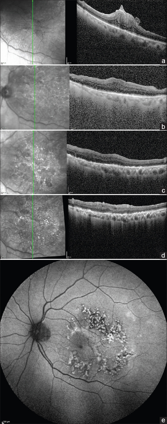
Purpose: To report a rare case of macular outer retinal and retinal pigment epithelium (RPE) damage following brilliant blue G (BBG)-assisted epiretinal membrane (ERM) removal surgery.
Methods: Retrospective, observational case report.
Results: An 85-year-old lady presented with decreased vision in the left eye and a best-corrected visual acuity of 20/400. The right eye examination was within normal limits. The left eye had a significant cataract, and the fundus examination through the cataractous haze showed an ERM with macular pucker, which was confirmed on an optical coherence tomography (OCT) scan. A combined cataract surgery with intraocular lens implantation and BBG-assisted ERM removal and internal limiting membrane peeling surgery was performed. Over the subsequent visits, a well-defined area of outer retinal and RPE alteration was identified on OCT and fundus autofluorescence without significant improvement in visual acuity. At the last follow-up visit, the visual acuity minimally improved to 20/200.
Conclusions: Macular toxicity due to repeated usage of BBG dye and high intensity focal endo-illumination may lead to poor visual outcome following ERM removal or similar macular surgeries. Adequate precautions need to be taken to prevent vision loss.
期刊介绍:
Peer Review under the responsibility of Iranian Society of Ophthalmology Journal of Current Ophthalmology, the official publication of the Iranian Society of Ophthalmology, is a peer-reviewed, open-access, scientific journal that welcomes high quality original articles related to vision science and all fields of ophthalmology. Journal of Current Ophthalmology is the continuum of Iranian Journal of Ophthalmology published since 1969.

 求助内容:
求助内容: 应助结果提醒方式:


