Kaleb Noruzi, Pooja Swami, Lidia Frejo, Jason Wright, Jason Wong, Daniel Grande, Timir Datta-Chaudhuri
{"title":"Effect of uniform capacitively coupled electric fields on matrix metabolism of osteoarthritic cartilage.","authors":"Kaleb Noruzi, Pooja Swami, Lidia Frejo, Jason Wright, Jason Wong, Daniel Grande, Timir Datta-Chaudhuri","doi":"10.1186/s42234-022-00096-w","DOIUrl":null,"url":null,"abstract":"<p><strong>Background: </strong>Osteoarthritis (OA) is a common and debilitating condition characterized by degeneration of hyaline cartilage. Currently, there is no treatment for OA that directly targets degradation of cartilage matrix. Capacitively coupled electric fields (CCEFs) represent a noninvasive and cost-effective treatment modality that can potentially restore articular cartilage homeostasis. Previous studies showed that stimulation of articular cartilage with CCEFs resulted in upregulation of anabolic factors and downregulation of catabolic factors. These studies didn't explain the derivation of the CCEFs or verify their uniformity and field strength, so it's possible that cartilage wasn't exposed to uniform field strength. The present study aims to employ CCEFs with verified uniform field strength in two in-vitro models of OA to investigate its potential to preserve cartilage matrix and validate the results of the aforementioned studies.</p><p><strong>Methods: </strong>Rabbit hyaline chondrocytes and full-thickness bovine articular cartilage explants were cultured in the absence or presence of CCEF and in the absence or presence of Interleukin1-B (IL-1B). Quantitative polymerase chain reaction (QPCR) was performed on chondrocytes to measure gene expression of ADAM-TS4, MMP3, MMP9, IL-6, TIMP1, and TIMP2. QPCR was performed on explants to measure gene expression of MMP3, Aggrecan, Collagen-2, and TIMP1. Aggrecan concentration in explants was measured with histology. Statistical analysis was performed using one-way analysis of variance and Tukey-Kramer multiple comparison test.</p><p><strong>Results: </strong>The treatment of chondrocytes with IL-1B resulted in upregulated expression of ADAM-TS4, MMP3, MMP9, and IL-6, while simultaneous administration of IL-1B and CCEF led to a relative decrease in ADAM-TS4, MMP3, MMP9, and IL-6 expression and a relative increase in TIMP1 and TIMP2 expression. Application of IL-1B and CCEF to the explants resulted in decreased expression of MMP3 and increased expression of Aggrecan, Collagen-2, and TIMP1 when compared to application of IL-1B alone.</p><p><strong>Conclusion: </strong>The data indicate that application of a CCEF with verified uniformity may result in upregulation of cartilage anabolic factors even in the presence of IL-1B while attenuating IL-1B induced upregulation of catabolic factors in both monolayer culture and whole tissue. These results demonstrate the potential of CCEFs to suppress the progression of OA and regenerate articular cartilage matrix.</p>","PeriodicalId":72363,"journal":{"name":"Bioelectronic medicine","volume":" ","pages":"14"},"PeriodicalIF":0.0000,"publicationDate":"2022-09-14","publicationTypes":"Journal Article","fieldsOfStudy":null,"isOpenAccess":false,"openAccessPdf":"https://www.ncbi.nlm.nih.gov/pmc/articles/PMC9472391/pdf/","citationCount":"0","resultStr":null,"platform":"Semanticscholar","paperid":null,"PeriodicalName":"Bioelectronic medicine","FirstCategoryId":"1085","ListUrlMain":"https://doi.org/10.1186/s42234-022-00096-w","RegionNum":0,"RegionCategory":null,"ArticlePicture":[],"TitleCN":null,"AbstractTextCN":null,"PMCID":null,"EPubDate":"","PubModel":"","JCR":"","JCRName":"","Score":null,"Total":0}
引用次数: 0
Abstract
Background: Osteoarthritis (OA) is a common and debilitating condition characterized by degeneration of hyaline cartilage. Currently, there is no treatment for OA that directly targets degradation of cartilage matrix. Capacitively coupled electric fields (CCEFs) represent a noninvasive and cost-effective treatment modality that can potentially restore articular cartilage homeostasis. Previous studies showed that stimulation of articular cartilage with CCEFs resulted in upregulation of anabolic factors and downregulation of catabolic factors. These studies didn't explain the derivation of the CCEFs or verify their uniformity and field strength, so it's possible that cartilage wasn't exposed to uniform field strength. The present study aims to employ CCEFs with verified uniform field strength in two in-vitro models of OA to investigate its potential to preserve cartilage matrix and validate the results of the aforementioned studies.
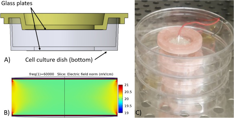
Methods: Rabbit hyaline chondrocytes and full-thickness bovine articular cartilage explants were cultured in the absence or presence of CCEF and in the absence or presence of Interleukin1-B (IL-1B). Quantitative polymerase chain reaction (QPCR) was performed on chondrocytes to measure gene expression of ADAM-TS4, MMP3, MMP9, IL-6, TIMP1, and TIMP2. QPCR was performed on explants to measure gene expression of MMP3, Aggrecan, Collagen-2, and TIMP1. Aggrecan concentration in explants was measured with histology. Statistical analysis was performed using one-way analysis of variance and Tukey-Kramer multiple comparison test.
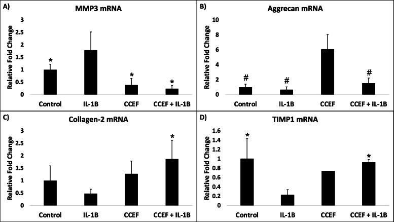
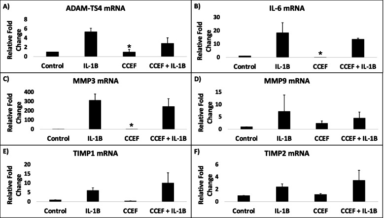
Results: The treatment of chondrocytes with IL-1B resulted in upregulated expression of ADAM-TS4, MMP3, MMP9, and IL-6, while simultaneous administration of IL-1B and CCEF led to a relative decrease in ADAM-TS4, MMP3, MMP9, and IL-6 expression and a relative increase in TIMP1 and TIMP2 expression. Application of IL-1B and CCEF to the explants resulted in decreased expression of MMP3 and increased expression of Aggrecan, Collagen-2, and TIMP1 when compared to application of IL-1B alone.
Conclusion: The data indicate that application of a CCEF with verified uniformity may result in upregulation of cartilage anabolic factors even in the presence of IL-1B while attenuating IL-1B induced upregulation of catabolic factors in both monolayer culture and whole tissue. These results demonstrate the potential of CCEFs to suppress the progression of OA and regenerate articular cartilage matrix.

 求助内容:
求助内容: 应助结果提醒方式:


