Alexandra Papoudou-Bai, Alexandra Barbouti, Vassiliki Galani, Kalliopi Stefanaki, Panagiotis Kanavaros
{"title":"Immunohistological analysis of the jun family and the signal transducers and activators of transcription in thymus.","authors":"Alexandra Papoudou-Bai, Alexandra Barbouti, Vassiliki Galani, Kalliopi Stefanaki, Panagiotis Kanavaros","doi":"10.1155/2015/541582","DOIUrl":null,"url":null,"abstract":"<p><p>The Jun family and the signal transducers and activators of transcription (STAT) are involved in proliferation and apoptosis. Moreover, c-Jun and STAT3 cooperate to regulate apoptosis. Therefore, we used double immunostaining to investigate the immunotopographical distribution of phospho-c-Jun (p-c-Jun), JunB, JunD, p-STAT3, p-STAT5, and p-STAT6 in human thymus. JunD was frequently expressed by thymocytes with higher expression in medullary compared to cortical thymocytes. p-c-Jun was frequently expressed by cortical and medullary thymic epithelial cells (TEC) and Hassall bodies (HB). p-STAT3 was frequently expressed by TEC with higher expression in cortical compared to medullary TEC and HB. p-c-Jun, JunB, p-STAT3, p-STAT5, and p-STAT6 were rarely expressed by thymocytes. JunB and JunD were expressed by rare cortical TEC with higher expression in medullary TEC. p-STAT5 and p-STAT6 were expressed by rare cortical and medullary TEC. Double immunostaining revealed p-c-Jun and JunD expression in rare CD11c positive dendritic cells. Our findings suggest a notable implication of JunD in the physiology of thymocytes and p-c-Jun and p-STAT3 in the physiology of TEC. The diversity of the immunotopographical distribution and the expression levels of p-c-Jun, JunB, JunD, p-STAT3, p-STAT5, and p-STAT6 indicates that they are differentially involved in the differentiation of TEC, thymocytes, and dendritic cells. </p>","PeriodicalId":89526,"journal":{"name":"Anatomy research international","volume":"2015 ","pages":"541582"},"PeriodicalIF":0.0000,"publicationDate":"2015-01-01","publicationTypes":"Journal Article","fieldsOfStudy":null,"isOpenAccess":false,"openAccessPdf":"https://www.ncbi.nlm.nih.gov/pmc/articles/PMC4381968/pdf/","citationCount":"2","resultStr":null,"platform":"Semanticscholar","paperid":null,"PeriodicalName":"Anatomy research international","FirstCategoryId":"1085","ListUrlMain":"https://doi.org/10.1155/2015/541582","RegionNum":0,"RegionCategory":null,"ArticlePicture":[],"TitleCN":null,"AbstractTextCN":null,"PMCID":null,"EPubDate":"2015/3/18 0:00:00","PubModel":"Epub","JCR":"","JCRName":"","Score":null,"Total":0}
引用次数: 2
Abstract
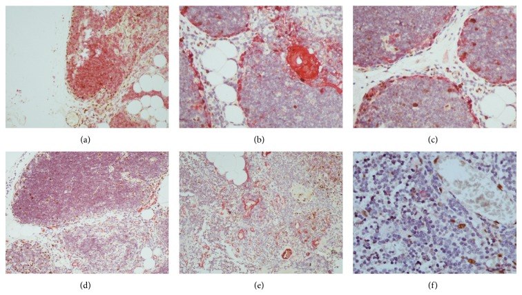
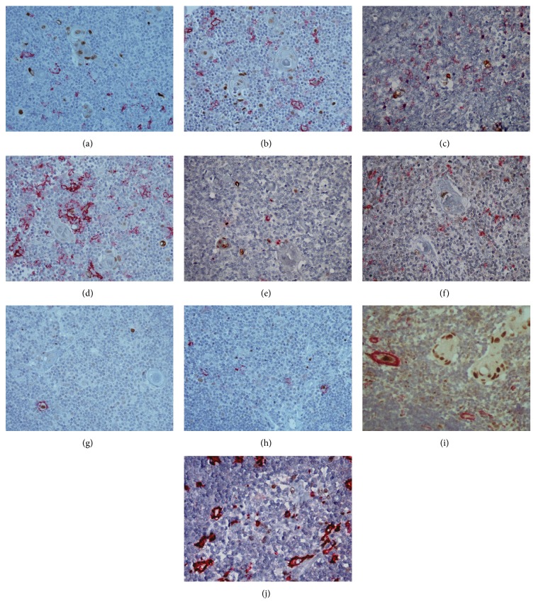
The Jun family and the signal transducers and activators of transcription (STAT) are involved in proliferation and apoptosis. Moreover, c-Jun and STAT3 cooperate to regulate apoptosis. Therefore, we used double immunostaining to investigate the immunotopographical distribution of phospho-c-Jun (p-c-Jun), JunB, JunD, p-STAT3, p-STAT5, and p-STAT6 in human thymus. JunD was frequently expressed by thymocytes with higher expression in medullary compared to cortical thymocytes. p-c-Jun was frequently expressed by cortical and medullary thymic epithelial cells (TEC) and Hassall bodies (HB). p-STAT3 was frequently expressed by TEC with higher expression in cortical compared to medullary TEC and HB. p-c-Jun, JunB, p-STAT3, p-STAT5, and p-STAT6 were rarely expressed by thymocytes. JunB and JunD were expressed by rare cortical TEC with higher expression in medullary TEC. p-STAT5 and p-STAT6 were expressed by rare cortical and medullary TEC. Double immunostaining revealed p-c-Jun and JunD expression in rare CD11c positive dendritic cells. Our findings suggest a notable implication of JunD in the physiology of thymocytes and p-c-Jun and p-STAT3 in the physiology of TEC. The diversity of the immunotopographical distribution and the expression levels of p-c-Jun, JunB, JunD, p-STAT3, p-STAT5, and p-STAT6 indicates that they are differentially involved in the differentiation of TEC, thymocytes, and dendritic cells.

 求助内容:
求助内容: 应助结果提醒方式:


