Marc Maegele, Ewa K Stuermer, Alexander Hoeffgen, Ulla Uhlenkueken, Angelika Mautes, Nadine Schaefer, Marcela Lippert-Gruener, Ute Schaefer, Mathias Hoehn
{"title":"Multimodal MR imaging of acute and subacute experimental traumatic brain injury: Time course and correlation with cerebral energy metabolites.","authors":"Marc Maegele, Ewa K Stuermer, Alexander Hoeffgen, Ulla Uhlenkueken, Angelika Mautes, Nadine Schaefer, Marcela Lippert-Gruener, Ute Schaefer, Mathias Hoehn","doi":"10.1177/2047981614555142","DOIUrl":null,"url":null,"abstract":"<p><strong>Background: </strong>Traumatic brain injury (TBI) is one of the leading causes of death and permanent disability world-wide. The predominant cause of death after TBI is brain edema which can be quantified by non-invasive diffusion-weighted magnetic resonance imaging (DWI).</p><p><strong>Purpose: </strong>To provide a better understanding of the early onset, time course, spatial development, and type of brain edema after TBI and to correlate MRI data and the cerebral energy state reflected by the metabolite adenosine triphosphate (ATP).</p><p><strong>Material and methods: </strong>The spontaneous development of lateral fluid percussion-induced TBI was investigated in the acute (6 h), subacute (48 h), and chronic (7 days) phase in rats by MRI of quantitative T2 and apparent diffusion coefficient (ADC) mapping as well as perfusion was combined with ATP-specific bioluminescence imaging and histology.</p><p><strong>Results: </strong>An induced TBI led to moderate to mild brain damages, reflected by transient, pronounced development of vasogenic edema and perfusion reduction. Heterogeneous ADC patterns indicated a parallel, but mixed expression of vasogenic and cytotoxic edema. Cortical ATP levels were reduced in the acute and subacute phase by 13% and 27%, respectively, but were completely normalized at 7 days after injury.</p><p><strong>Conclusion: </strong>The partial ATP reduction was interpreted to be partially caused by a loss of neurons in parallel with transient dilution of the regional ATP concentration by pronounced vasogenic edema. The normalization of energy metabolism after 7 days was likely due to infiltrating glia and not to recovery. The MRI combined with metabolite measurement further improves the understanding and evaluation of brain damages after TBI.</p>","PeriodicalId":30445,"journal":{"name":"Acta Radiologica Short Reports","volume":"4 1","pages":"2047981614555142"},"PeriodicalIF":0.0000,"publicationDate":"2015-01-06","publicationTypes":"Journal Article","fieldsOfStudy":null,"isOpenAccess":false,"openAccessPdf":"https://ftp.ncbi.nlm.nih.gov/pub/pmc/oa_pdf/61/d7/10.1177_2047981614555142.PMC4299368.pdf","citationCount":"0","resultStr":null,"platform":"Semanticscholar","paperid":null,"PeriodicalName":"Acta Radiologica Short Reports","FirstCategoryId":"1085","ListUrlMain":"https://doi.org/10.1177/2047981614555142","RegionNum":0,"RegionCategory":null,"ArticlePicture":[],"TitleCN":null,"AbstractTextCN":null,"PMCID":null,"EPubDate":"2015/1/1 0:00:00","PubModel":"eCollection","JCR":"","JCRName":"","Score":null,"Total":0}
引用次数: 0
Abstract
Background: Traumatic brain injury (TBI) is one of the leading causes of death and permanent disability world-wide. The predominant cause of death after TBI is brain edema which can be quantified by non-invasive diffusion-weighted magnetic resonance imaging (DWI).
Purpose: To provide a better understanding of the early onset, time course, spatial development, and type of brain edema after TBI and to correlate MRI data and the cerebral energy state reflected by the metabolite adenosine triphosphate (ATP).
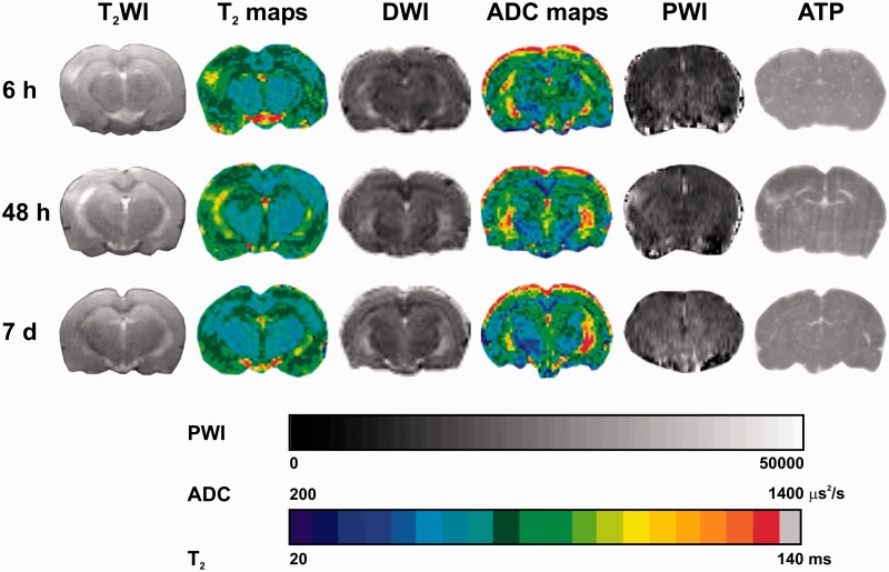
Material and methods: The spontaneous development of lateral fluid percussion-induced TBI was investigated in the acute (6 h), subacute (48 h), and chronic (7 days) phase in rats by MRI of quantitative T2 and apparent diffusion coefficient (ADC) mapping as well as perfusion was combined with ATP-specific bioluminescence imaging and histology.
Results: An induced TBI led to moderate to mild brain damages, reflected by transient, pronounced development of vasogenic edema and perfusion reduction. Heterogeneous ADC patterns indicated a parallel, but mixed expression of vasogenic and cytotoxic edema. Cortical ATP levels were reduced in the acute and subacute phase by 13% and 27%, respectively, but were completely normalized at 7 days after injury.
Conclusion: The partial ATP reduction was interpreted to be partially caused by a loss of neurons in parallel with transient dilution of the regional ATP concentration by pronounced vasogenic edema. The normalization of energy metabolism after 7 days was likely due to infiltrating glia and not to recovery. The MRI combined with metabolite measurement further improves the understanding and evaluation of brain damages after TBI.
背景:创伤性脑损伤(TBI)是导致全球死亡和永久性残疾的主要原因之一。目的:更好地了解创伤性脑损伤后脑水肿的早期发生、时间进程、空间发展和类型,并将磁共振成像数据与代谢物三磷酸腺苷(ATP)反映的脑能量状态相关联:通过核磁共振成像的定量T2和表观扩散系数(ADC)映射以及灌注,并结合ATP特异性生物发光成像和组织学,研究了大鼠在急性期(6小时)、亚急性期(48小时)和慢性期(7天)自发发展的外侧流体叩击诱导的创伤性脑损伤:结果:诱导性创伤性脑损伤导致中度至轻度脑损伤,表现为短暂、明显的血管源性水肿和灌注减少。不同的 ADC 模式表明血管源性水肿和细胞毒性水肿并存,但表现不一。皮质 ATP 水平在急性期和亚急性期分别降低了 13% 和 27%,但在损伤后 7 天完全恢复正常:部分 ATP 减少的部分原因是神经元丢失,同时明显的血管源性水肿造成区域 ATP 浓度短暂稀释。7天后能量代谢恢复正常可能是由于胶质细胞的浸润,而非恢复所致。核磁共振成像与代谢物测量相结合,进一步提高了对创伤性脑损伤后脑损伤的认识和评估。
期刊介绍:
Under the editorial leadership of Professor Arnulf Skjennald MD and a distinguished editorial board, Acta Radiologica Open, formerly Acta Radiologica Short Reports, aims for the prompt publication of original case reports, short reports, review articles, pictorial reviews, research articles on diagnostic and interventional radiology, clinical radiology, experimental investigations in animals, and all other research related to imaging procedures. Acta Radiologica Open provides a complete update on all radiological specialties and technical utilities, as well as physiology and physics related to imaging, including ultrasonography, computed tomography, radionuclide and magnetic resonance imaging. Acta Radiologica Open publishes articles on diagnostic and interventional procedures in radiology based on all medical imaging techniques, as well as works in physiology and physics when related to radiology. The journal is an online-only, peer reviewed, open access journal.


 求助内容:
求助内容: 应助结果提醒方式:


