Khoschy Schawkat, Beatrix Hoksch, Markus Schwerzmann, Stefan Puig, Thorsten Klink
{"title":"Diagnosis of cardiac metastasis from cervical cancer in a 33-year-old patient using multimodal imaging studies: a case report and literature review.","authors":"Khoschy Schawkat, Beatrix Hoksch, Markus Schwerzmann, Stefan Puig, Thorsten Klink","doi":"10.1177/2047981614530287","DOIUrl":null,"url":null,"abstract":"<p><p>We report a case of a 33-year-old woman with emergency admission due to dyspnoea and fever. History included squamous cell carcinoma of the cervix in complete remission. Contrast-enhanced computed tomography (CT) scanning of the chest, which was indicated to rule out pneumonia, revealed an infiltrative cardiac mass. Further assessment of the tumour by echocardiography and cardiac magnetic resonance imaging (MRI) showed transmural infiltration of the apical interventricular septum with a mass extending into the left and right ventricle cavities. The mass was highly suspicious for a cardiac metastasis. Cardiac metastases from cervical cancer are extremely rare. Recurrence of cervical carcinoma involving the heart should be considered even after a curative therapy approach. Non-invasive imaging plays a paramount role in investigating cardiac masses. Echocardiography, CT and MRI are complementary imaging modalities for complete work-up of intracardiac lesions. </p>","PeriodicalId":30445,"journal":{"name":"Acta Radiologica Short Reports","volume":"3 8","pages":"2047981614530287"},"PeriodicalIF":0.0000,"publicationDate":"2014-09-15","publicationTypes":"Journal Article","fieldsOfStudy":null,"isOpenAccess":false,"openAccessPdf":"https://sci-hub-pdf.com/10.1177/2047981614530287","citationCount":"8","resultStr":null,"platform":"Semanticscholar","paperid":null,"PeriodicalName":"Acta Radiologica Short Reports","FirstCategoryId":"1085","ListUrlMain":"https://doi.org/10.1177/2047981614530287","RegionNum":0,"RegionCategory":null,"ArticlePicture":[],"TitleCN":null,"AbstractTextCN":null,"PMCID":null,"EPubDate":"2014/9/1 0:00:00","PubModel":"eCollection","JCR":"","JCRName":"","Score":null,"Total":0}
引用次数: 8
Abstract
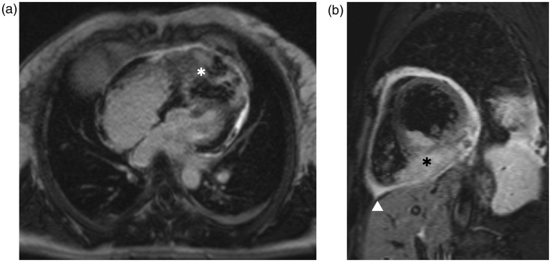
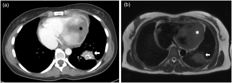
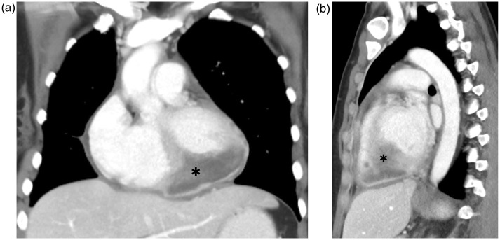
We report a case of a 33-year-old woman with emergency admission due to dyspnoea and fever. History included squamous cell carcinoma of the cervix in complete remission. Contrast-enhanced computed tomography (CT) scanning of the chest, which was indicated to rule out pneumonia, revealed an infiltrative cardiac mass. Further assessment of the tumour by echocardiography and cardiac magnetic resonance imaging (MRI) showed transmural infiltration of the apical interventricular septum with a mass extending into the left and right ventricle cavities. The mass was highly suspicious for a cardiac metastasis. Cardiac metastases from cervical cancer are extremely rare. Recurrence of cervical carcinoma involving the heart should be considered even after a curative therapy approach. Non-invasive imaging plays a paramount role in investigating cardiac masses. Echocardiography, CT and MRI are complementary imaging modalities for complete work-up of intracardiac lesions.
期刊介绍:
Under the editorial leadership of Professor Arnulf Skjennald MD and a distinguished editorial board, Acta Radiologica Open, formerly Acta Radiologica Short Reports, aims for the prompt publication of original case reports, short reports, review articles, pictorial reviews, research articles on diagnostic and interventional radiology, clinical radiology, experimental investigations in animals, and all other research related to imaging procedures. Acta Radiologica Open provides a complete update on all radiological specialties and technical utilities, as well as physiology and physics related to imaging, including ultrasonography, computed tomography, radionuclide and magnetic resonance imaging. Acta Radiologica Open publishes articles on diagnostic and interventional procedures in radiology based on all medical imaging techniques, as well as works in physiology and physics when related to radiology. The journal is an online-only, peer reviewed, open access journal.

 求助内容:
求助内容: 应助结果提醒方式:


