Joanna J Moser, Marvin J Fritzler, Jerome B Rattner
{"title":"Ultrastructural characterization of primary cilia in pathologically characterized human glioblastoma multiforme (GBM) tumors.","authors":"Joanna J Moser, Marvin J Fritzler, Jerome B Rattner","doi":"10.1186/1472-6890-14-40","DOIUrl":null,"url":null,"abstract":"<p><strong>Background: </strong>Primary cilia are non-motile sensory cytoplasmic organelles that are involved in cell cycle progression. Ultrastructurally, the primary cilium region is complex, with normal ciliogenesis progressing through five distinct morphological stages in human astrocytes. Defects in early stages of ciliogenesis are key features of astrocytoma/glioblastoma cell lines and provided the impetus for the current study which describes the morphology of primary cilia in molecularly characterized human glioblastoma multiforme (GBM) tumors.</p><p><strong>Methods: </strong>Seven surgically resected human GBM tissue samples were molecularly characterized according to IDH1/2 mutation status, EGFR amplification status and MGMT promoter methylation status and were examined for primary cilia expression and structure using indirect immunofluorescence and electron microscopy.</p><p><strong>Results: </strong>We report for the first time that primary cilia are disrupted in the early stages of ciliogenesis in human GBM tumors. We confirm that immature primary cilia and basal bodies/centrioles have aberrant ciliogenesis characteristics including absent paired vesicles, misshaped/swollen vesicular hats, abnormal configuration of distal appendages, and discontinuity of centriole microtubular blades. Additionally, the transition zone plate is able to form in the absence of paired vesicles on the distal end of the basal body and when a cilium progresses beyond the early stages of ciliogenesis, it has electron dense material clumped along the transition zone and a darkening of the microtubules at the proximal end of the cilium.</p><p><strong>Conclusions: </strong>Primary cilia play a role in a variety of human cancers. Previously primary cilia structure was perturbed in cultured cell lines derived from astrocytomas/glioblastomas; however there was always some question as to whether these findings were a cell culture phenomena. In this study we confirm that disruptions in ciliogenesis at early stages do occur in GBM tumors and that these ultrastructural findings bear resemblance to those previously observed in cell cultures. This is the first study to demonstrate that defects in cilia expression and function are a true hallmark of GBM tumors and correlate with their unrestrained growth. A review of the current ultrastructural profiles in the literature provides suggestions as to the best possible candidate protein that underlies defects in the early stages of ciliogenesis within GBM tumors.</p>","PeriodicalId":35804,"journal":{"name":"BMC Clinical Pathology","volume":"14 ","pages":"40"},"PeriodicalIF":0.0000,"publicationDate":"2014-09-12","publicationTypes":"Journal Article","fieldsOfStudy":null,"isOpenAccess":false,"openAccessPdf":"https://sci-hub-pdf.com/10.1186/1472-6890-14-40","citationCount":"41","resultStr":null,"platform":"Semanticscholar","paperid":null,"PeriodicalName":"BMC Clinical Pathology","FirstCategoryId":"1085","ListUrlMain":"https://doi.org/10.1186/1472-6890-14-40","RegionNum":0,"RegionCategory":null,"ArticlePicture":[],"TitleCN":null,"AbstractTextCN":null,"PMCID":null,"EPubDate":"2014/1/1 0:00:00","PubModel":"eCollection","JCR":"Q2","JCRName":"Medicine","Score":null,"Total":0}
引用次数: 41
Abstract
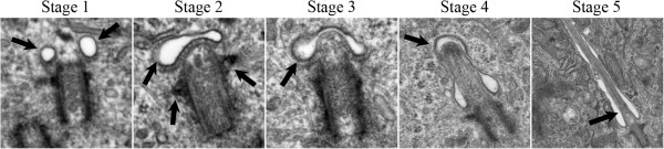
Background: Primary cilia are non-motile sensory cytoplasmic organelles that are involved in cell cycle progression. Ultrastructurally, the primary cilium region is complex, with normal ciliogenesis progressing through five distinct morphological stages in human astrocytes. Defects in early stages of ciliogenesis are key features of astrocytoma/glioblastoma cell lines and provided the impetus for the current study which describes the morphology of primary cilia in molecularly characterized human glioblastoma multiforme (GBM) tumors.
Methods: Seven surgically resected human GBM tissue samples were molecularly characterized according to IDH1/2 mutation status, EGFR amplification status and MGMT promoter methylation status and were examined for primary cilia expression and structure using indirect immunofluorescence and electron microscopy.
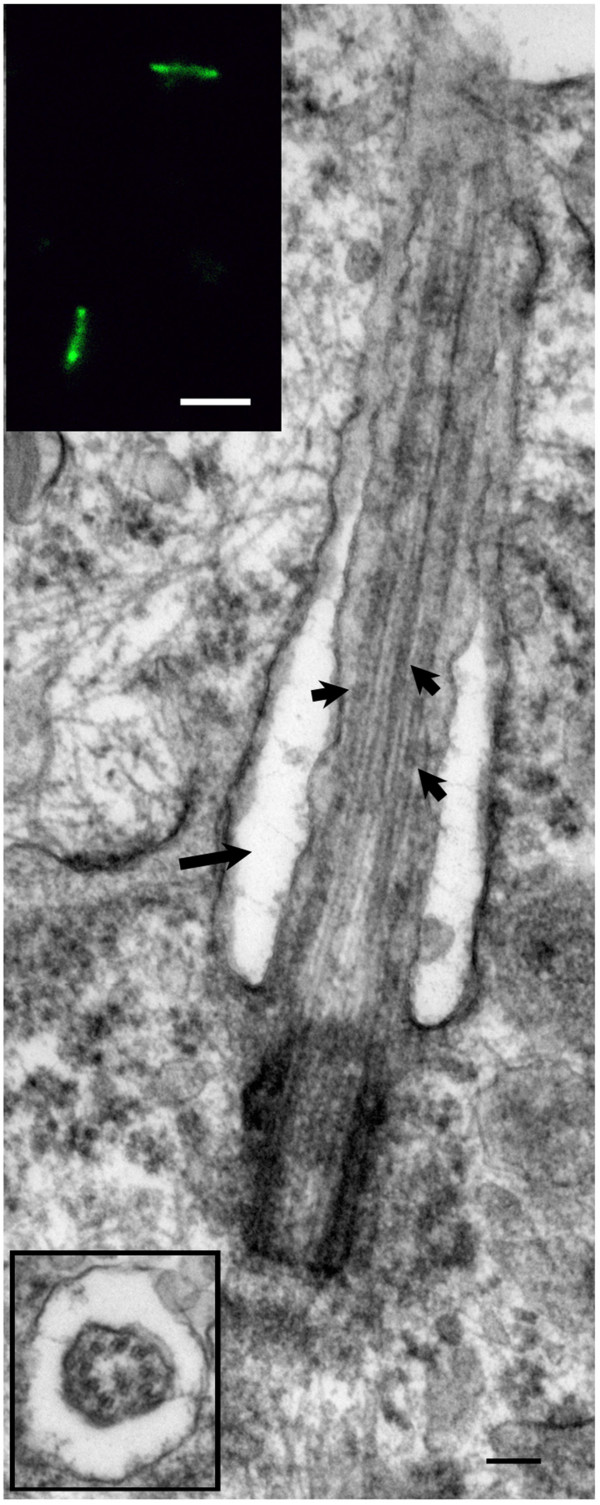
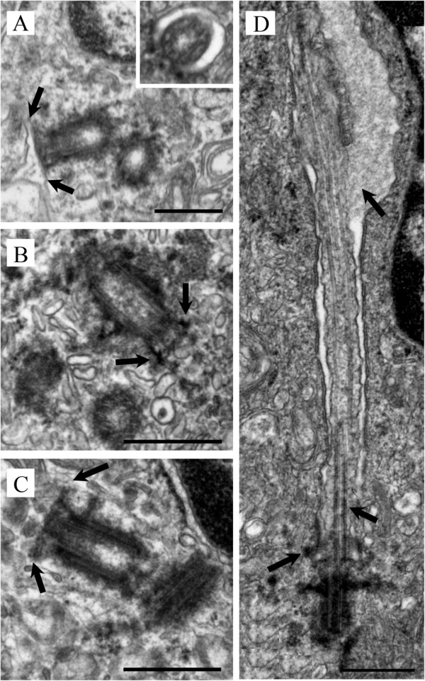
Results: We report for the first time that primary cilia are disrupted in the early stages of ciliogenesis in human GBM tumors. We confirm that immature primary cilia and basal bodies/centrioles have aberrant ciliogenesis characteristics including absent paired vesicles, misshaped/swollen vesicular hats, abnormal configuration of distal appendages, and discontinuity of centriole microtubular blades. Additionally, the transition zone plate is able to form in the absence of paired vesicles on the distal end of the basal body and when a cilium progresses beyond the early stages of ciliogenesis, it has electron dense material clumped along the transition zone and a darkening of the microtubules at the proximal end of the cilium.
Conclusions: Primary cilia play a role in a variety of human cancers. Previously primary cilia structure was perturbed in cultured cell lines derived from astrocytomas/glioblastomas; however there was always some question as to whether these findings were a cell culture phenomena. In this study we confirm that disruptions in ciliogenesis at early stages do occur in GBM tumors and that these ultrastructural findings bear resemblance to those previously observed in cell cultures. This is the first study to demonstrate that defects in cilia expression and function are a true hallmark of GBM tumors and correlate with their unrestrained growth. A review of the current ultrastructural profiles in the literature provides suggestions as to the best possible candidate protein that underlies defects in the early stages of ciliogenesis within GBM tumors.
期刊介绍:
BMC Clinical Pathology is an open access journal publishing original peer-reviewed research articles in all aspects of histopathology, haematology, clinical biochemistry, and medical microbiology (including virology, parasitology, and infection control). BMC Clinical Pathology (ISSN 1472-6890) is indexed/tracked/covered by PubMed, CAS, EMBASE, Scopus and Google Scholar.

 求助内容:
求助内容: 应助结果提醒方式:


