Chordoid meningioma in a pediatric patient with tuberous sclerosis complex.

Korean Journal of Pathology Pub Date : 2014-08-01 Epub Date: 2014-08-26 DOI:10.4132/KoreanJPathol.2014.48.4.302
Jiwon Lee, Hee Joon Yu, Jeehun Lee, Ji Hye Kim, Hyung Jin Shin, Yeon-Lim Suh, Munhyang Lee
{"title":"Chordoid meningioma in a pediatric patient with tuberous sclerosis complex.","authors":"Jiwon Lee, Hee Joon Yu, Jeehun Lee, Ji Hye Kim, Hyung Jin Shin, Yeon-Lim Suh, Munhyang Lee","doi":"10.4132/KoreanJPathol.2014.48.4.302","DOIUrl":null,"url":null,"abstract":"Meningeal tumors are rare in childhood, comprising less than 4.2% of all pediatric primary intracranial tumors.1 Fourteen histological subtypes of meningiomas exist in the World Health Organization (WHO) classification system.2 Chordoid meningioma (CM) is a rare meningiomal variant characterized histologically by features that mimic chordoma and other chordoid neoplasms.2 Chordoid meningiomal tumors tend to exhibit local recurrence and aggressive behavior, although they typically show morphologically benign features. \n \nIn a tuberous sclerosis complex (TSC) patient, hamartomas can occur in multiple organs and cause diverse clinical symptoms. Cortical tubers, subependymal nodules (SENs) and subependymal giant cell astrocytomas (SEGAs) are representative lesions of TSC and can be detected by magnetic resonance imaging (MRI) of the brain.3 SEGA lesions have been found to occur in approximately 6.1% to 18.5% of TSC patients, and account for 90% of all intracranial tumors associated with TSC.4 SEGA lesions are commonly located in the foramen of Monro or the periventricular region,5 and can cause loss of vision, papillary edemas, intracranial calcification, and hydrocephalus.6 The primary method for treating SEGA lesions consists of surgical resection of the tumors; complete and early surgical removal of tumors has been shown to be the most important prognostic factor.6 \n \nChildhood CM has been shown to account for only ~0.5-1% of all meningiomas, with intraventricular occurrence of CM reported in only 27 children to date.2,7 Also, CM has not been reported in any TSC patients to date. Here we report a case of intraventricular CM, which appeared highly similar to SEGA in brain MRI scans, in a child with TSC.","PeriodicalId":49936,"journal":{"name":"Korean Journal of Pathology","volume":"48 4","pages":"302-6"},"PeriodicalIF":0.0000,"publicationDate":"2014-08-01","publicationTypes":"Journal Article","fieldsOfStudy":null,"isOpenAccess":false,"openAccessPdf":"https://sci-hub-pdf.com/10.4132/KoreanJPathol.2014.48.4.302","citationCount":"4","resultStr":null,"platform":"Semanticscholar","paperid":null,"PeriodicalName":"Korean Journal of Pathology","FirstCategoryId":"1085","ListUrlMain":"https://doi.org/10.4132/KoreanJPathol.2014.48.4.302","RegionNum":0,"RegionCategory":null,"ArticlePicture":[],"TitleCN":null,"AbstractTextCN":null,"PMCID":null,"EPubDate":"2014/8/26 0:00:00","PubModel":"Epub","JCR":"","JCRName":"","Score":null,"Total":0}
引用次数: 4

Abstract

Meningeal tumors are rare in childhood, comprising less than 4.2% of all pediatric primary intracranial tumors.1 Fourteen histological subtypes of meningiomas exist in the World Health Organization (WHO) classification system.2 Chordoid meningioma (CM) is a rare meningiomal variant characterized histologically by features that mimic chordoma and other chordoid neoplasms.2 Chordoid meningiomal tumors tend to exhibit local recurrence and aggressive behavior, although they typically show morphologically benign features. In a tuberous sclerosis complex (TSC) patient, hamartomas can occur in multiple organs and cause diverse clinical symptoms. Cortical tubers, subependymal nodules (SENs) and subependymal giant cell astrocytomas (SEGAs) are representative lesions of TSC and can be detected by magnetic resonance imaging (MRI) of the brain.3 SEGA lesions have been found to occur in approximately 6.1% to 18.5% of TSC patients, and account for 90% of all intracranial tumors associated with TSC.4 SEGA lesions are commonly located in the foramen of Monro or the periventricular region,5 and can cause loss of vision, papillary edemas, intracranial calcification, and hydrocephalus.6 The primary method for treating SEGA lesions consists of surgical resection of the tumors; complete and early surgical removal of tumors has been shown to be the most important prognostic factor.6 Childhood CM has been shown to account for only ~0.5-1% of all meningiomas, with intraventricular occurrence of CM reported in only 27 children to date.2,7 Also, CM has not been reported in any TSC patients to date. Here we report a case of intraventricular CM, which appeared highly similar to SEGA in brain MRI scans, in a child with TSC.
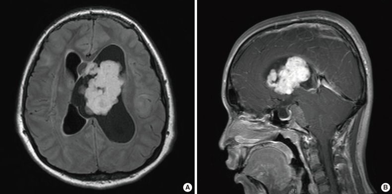
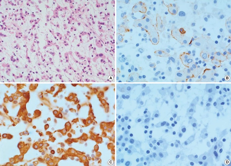
Abstract Image

Abstract Image

小儿结节性硬化症合并脊索样脑膜瘤1例。
本文章由计算机程序翻译,如有差异,请以英文原文为准。
求助全文
约1分钟内获得全文 求助全文
来源期刊
Korean Journal of Pathology
Korean Journal of Pathology 医学-病理学
自引率
0.00%
发文量
0
审稿时长
6-12 weeks
×
引用
GB/T 7714-2015
复制
MLA
复制
APA
复制
导出至
BibTeX EndNote RefMan NoteFirst NoteExpress
×
提示
您的信息不完整,为了账户安全,请先补充。
现在去补充
×
提示
您因"违规操作"
具体请查看互助需知
我知道了
×
提示
确定
请完成安全验证×
copy
已复制链接
快去分享给好友吧!
我知道了
右上角分享
点击右上角分享
0
联系我们:info@booksci.cn Book学术提供免费学术资源搜索服务,方便国内外学者检索中英文文献。致力于提供最便捷和优质的服务体验。 Copyright © 2023 布克学术 All rights reserved.
京ICP备2023020795号-1
ghs 京公网安备 11010802042870号
Book学术文献互助
Book学术文献互助群
群 号:481959085
Book学术官方微信