Hwa Jin Cho, Han Na Kim, Kyung Ju Kim, Kyu Sang Lee, Jae Kyung Myung, Seung-Ki Kim, Sung-Hye Park
{"title":"Intracranial extracerebral glioneuronal heterotopia with adipose tissue and a glioependymal cyst: a case report and review of the literature.","authors":"Hwa Jin Cho, Han Na Kim, Kyung Ju Kim, Kyu Sang Lee, Jae Kyung Myung, Seung-Ki Kim, Sung-Hye Park","doi":"10.4132/KoreanJPathol.2014.48.3.254","DOIUrl":null,"url":null,"abstract":"The presence of central nervous system (CNS) tissue outside the cranium is often referred to as \"heterotopia,\" although technically this should be termed \"ectopia,\" according to the dictionary definition. Glioneuronal heterotopia (GH) is a rare, mass-forming, malformative lesion. Ectopic glioneuronal tissue of the head and neck has been detected in the nasopharynx, oropharynx, tongue, palate, tonsils, soft tissue, eye, and orbit, and intracranial extracerebral glioneuronal heterotopia (IEGH) has also been reported, although less frequently.1,2 Since the first description of neuroglial heterotopia in the dorsal meninges of the cervical spinal cord by Wolbach in 1907,3 fewer than 20 cases of IEGH have been reported. Glioependymal cysts are rare, ependyma-lined, cystic lesions of the subarachnoid space, which have been referred to as epithelial or ependymal cysts. Histopathologically, they are lined with ependymal cells abutted on the glial layer and are commonly detected in the posterior fossa. The origin of glioependymal cysts of the posterior fossa is not clear, but these cysts may represent neuroglial heterotopia, persistent Blake's pouch (diverticulum of the roof of the fourth ventricle), or remnants of a tela chorioidea. We report here a case of IEGH that was predominantly composed of cerebellar tissue with some fat tissue and a large glioependymal cyst, and was initially misdiagnosed as a teratoma with a glioependymal cyst.","PeriodicalId":49936,"journal":{"name":"Korean Journal of Pathology","volume":"48 3","pages":"254-7"},"PeriodicalIF":0.0000,"publicationDate":"2014-06-01","publicationTypes":"Journal Article","fieldsOfStudy":null,"isOpenAccess":false,"openAccessPdf":"https://sci-hub-pdf.com/10.4132/KoreanJPathol.2014.48.3.254","citationCount":"5","resultStr":null,"platform":"Semanticscholar","paperid":null,"PeriodicalName":"Korean Journal of Pathology","FirstCategoryId":"1085","ListUrlMain":"https://doi.org/10.4132/KoreanJPathol.2014.48.3.254","RegionNum":0,"RegionCategory":null,"ArticlePicture":[],"TitleCN":null,"AbstractTextCN":null,"PMCID":null,"EPubDate":"2014/6/26 0:00:00","PubModel":"Epub","JCR":"","JCRName":"","Score":null,"Total":0}
引用次数: 5
Abstract
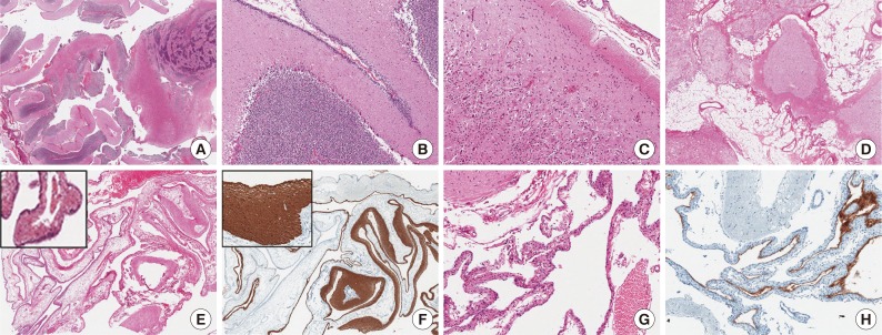
The presence of central nervous system (CNS) tissue outside the cranium is often referred to as "heterotopia," although technically this should be termed "ectopia," according to the dictionary definition. Glioneuronal heterotopia (GH) is a rare, mass-forming, malformative lesion. Ectopic glioneuronal tissue of the head and neck has been detected in the nasopharynx, oropharynx, tongue, palate, tonsils, soft tissue, eye, and orbit, and intracranial extracerebral glioneuronal heterotopia (IEGH) has also been reported, although less frequently.1,2 Since the first description of neuroglial heterotopia in the dorsal meninges of the cervical spinal cord by Wolbach in 1907,3 fewer than 20 cases of IEGH have been reported. Glioependymal cysts are rare, ependyma-lined, cystic lesions of the subarachnoid space, which have been referred to as epithelial or ependymal cysts. Histopathologically, they are lined with ependymal cells abutted on the glial layer and are commonly detected in the posterior fossa. The origin of glioependymal cysts of the posterior fossa is not clear, but these cysts may represent neuroglial heterotopia, persistent Blake's pouch (diverticulum of the roof of the fourth ventricle), or remnants of a tela chorioidea. We report here a case of IEGH that was predominantly composed of cerebellar tissue with some fat tissue and a large glioependymal cyst, and was initially misdiagnosed as a teratoma with a glioependymal cyst.