{"title":"Well-differentiated papillary mesothelioma of the tunica vaginalis: a case study and review of the literature.","authors":"Seyda Erdogan, Arbil Acikalin, Handan Zeren, Gulfılız Gonlusen, Suzan Zorludemir, Volkan Izol","doi":"10.4132/KoreanJPathol.2014.48.3.225","DOIUrl":null,"url":null,"abstract":"<p><p>Well-differentiated papillary mesothelioma is an uncommon tumor of the testes that usually presents as a hydrocele. Here, we present the case of one patient who did not have a history of asbestos exposure. The tumor was localized in the tunica vaginalis and was composed of three pedunculated masses macroscopically. Microscopically, branching papillary structures with focal coagulative necrosis were present. In addition to immunohistochemistry, simian virus 40 DNA was also tested by polymerase chain reaction. This report presents one case of this rare entity, its clinical and macroscopic features, and follow-up results. </p>","PeriodicalId":49936,"journal":{"name":"Korean Journal of Pathology","volume":"48 3","pages":"225-8"},"PeriodicalIF":0.0000,"publicationDate":"2014-06-01","publicationTypes":"Journal Article","fieldsOfStudy":null,"isOpenAccess":false,"openAccessPdf":"https://sci-hub-pdf.com/10.4132/KoreanJPathol.2014.48.3.225","citationCount":"15","resultStr":null,"platform":"Semanticscholar","paperid":null,"PeriodicalName":"Korean Journal of Pathology","FirstCategoryId":"1085","ListUrlMain":"https://doi.org/10.4132/KoreanJPathol.2014.48.3.225","RegionNum":0,"RegionCategory":null,"ArticlePicture":[],"TitleCN":null,"AbstractTextCN":null,"PMCID":null,"EPubDate":"2014/6/26 0:00:00","PubModel":"Epub","JCR":"","JCRName":"","Score":null,"Total":0}
引用次数: 15
Abstract
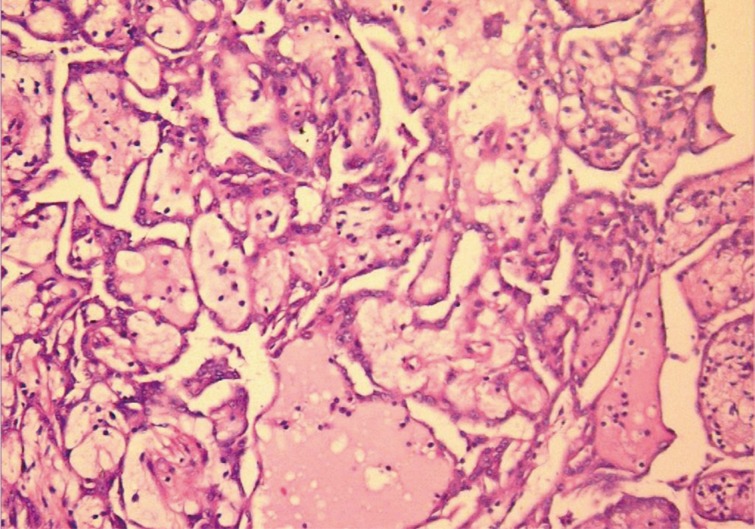
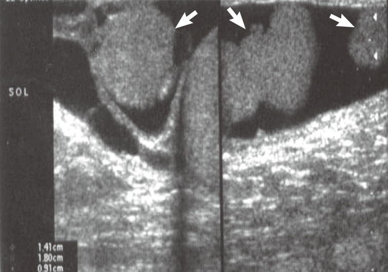
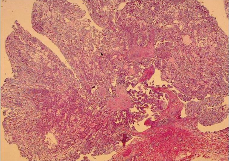
Well-differentiated papillary mesothelioma is an uncommon tumor of the testes that usually presents as a hydrocele. Here, we present the case of one patient who did not have a history of asbestos exposure. The tumor was localized in the tunica vaginalis and was composed of three pedunculated masses macroscopically. Microscopically, branching papillary structures with focal coagulative necrosis were present. In addition to immunohistochemistry, simian virus 40 DNA was also tested by polymerase chain reaction. This report presents one case of this rare entity, its clinical and macroscopic features, and follow-up results.