{"title":"Vascular waveform analysis of flap-feeding vessels using color Doppler ultrasonography.","authors":"Akihiro Ogino, Kiyoshi Onishi","doi":"10.1155/2014/249670","DOIUrl":null,"url":null,"abstract":"<p><p>We performed vascular waveform analysis of flap-feeding vessels using color Doppler ultrasonography and evaluated the blood flow in the flaps prior to surgery. Vascular waveform analysis was performed in 19 patients. The analyzed parameters included the vascular diameter, flow volume, flow velocity, resistance index, pulsatility index, and acceleration time. The arterial waveform was classified into 5 types based on the partially modified blood flow waveform classification reported by Hirai et al.; in particular, D-1a, D-1b, and D-2 were considered as normal waveforms. They were 4 patients which observed abnormal vascular waveform among 19 patients (D-4 : 1, D-3 : 1, and Poor detect : 2). The case which presented D-4 waveform changed the surgical procedure, and a favorable outcome was achieved. Muscle flap of the case which presented D-3 waveform was partially necrosed. The case which detected blood flow poorly was judged to be the vascular obstruction of the internal thoracic artery. In the evaluation of blood flow in flaps using color Doppler ultrasonography, determination of not only basic blood flow information, such as the vascular distribution and diameter and flow velocity, but also the flow volume, vascular resistance, and arterial waveform is essential to elucidate the hemodynamics of the flap. </p>","PeriodicalId":20105,"journal":{"name":"Plastic Surgery International","volume":"2014 ","pages":"249670"},"PeriodicalIF":0.0000,"publicationDate":"2014-01-01","publicationTypes":"Journal Article","fieldsOfStudy":null,"isOpenAccess":false,"openAccessPdf":"https://sci-hub-pdf.com/10.1155/2014/249670","citationCount":"8","resultStr":null,"platform":"Semanticscholar","paperid":null,"PeriodicalName":"Plastic Surgery International","FirstCategoryId":"1085","ListUrlMain":"https://doi.org/10.1155/2014/249670","RegionNum":0,"RegionCategory":null,"ArticlePicture":[],"TitleCN":null,"AbstractTextCN":null,"PMCID":null,"EPubDate":"2014/4/7 0:00:00","PubModel":"Epub","JCR":"","JCRName":"","Score":null,"Total":0}
引用次数: 8
Abstract
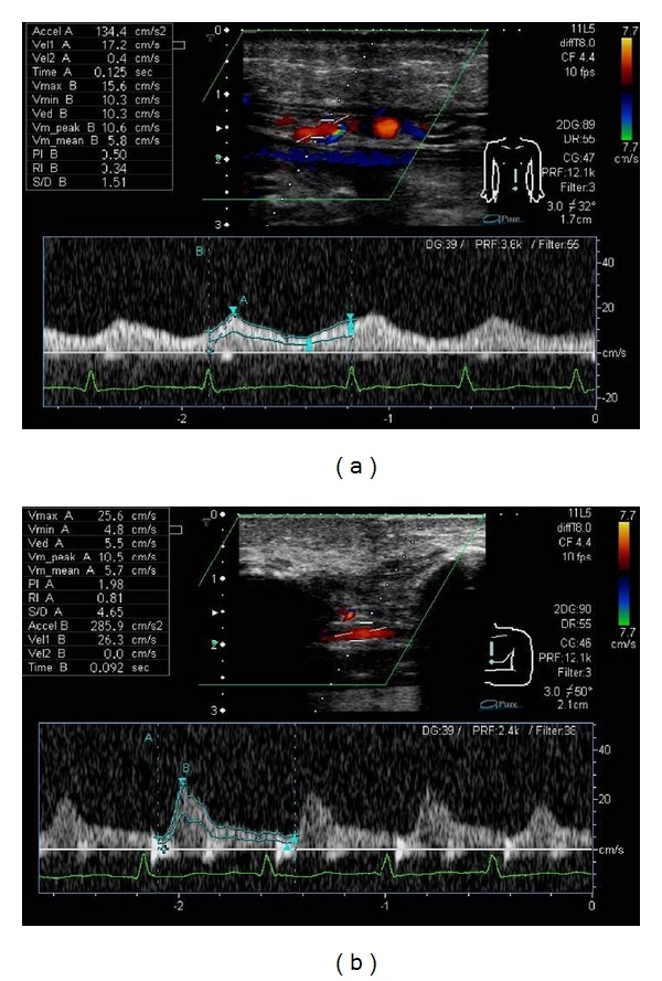
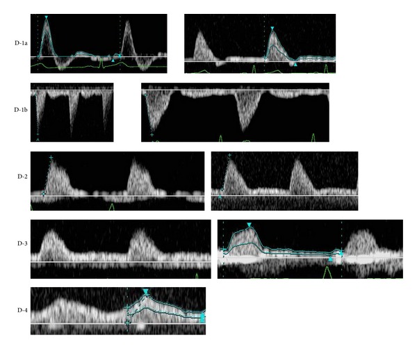
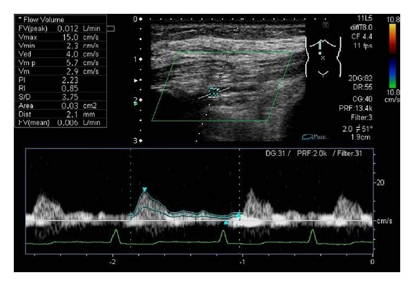
We performed vascular waveform analysis of flap-feeding vessels using color Doppler ultrasonography and evaluated the blood flow in the flaps prior to surgery. Vascular waveform analysis was performed in 19 patients. The analyzed parameters included the vascular diameter, flow volume, flow velocity, resistance index, pulsatility index, and acceleration time. The arterial waveform was classified into 5 types based on the partially modified blood flow waveform classification reported by Hirai et al.; in particular, D-1a, D-1b, and D-2 were considered as normal waveforms. They were 4 patients which observed abnormal vascular waveform among 19 patients (D-4 : 1, D-3 : 1, and Poor detect : 2). The case which presented D-4 waveform changed the surgical procedure, and a favorable outcome was achieved. Muscle flap of the case which presented D-3 waveform was partially necrosed. The case which detected blood flow poorly was judged to be the vascular obstruction of the internal thoracic artery. In the evaluation of blood flow in flaps using color Doppler ultrasonography, determination of not only basic blood flow information, such as the vascular distribution and diameter and flow velocity, but also the flow volume, vascular resistance, and arterial waveform is essential to elucidate the hemodynamics of the flap.

 求助内容:
求助内容: 应助结果提醒方式:


