Analysis by Light, Scanning, and Transmission Microscopy of the Intima Synovial of the Temporomandibular Joint of Human Fetuses during the Development.
Carlos Sabu Alvez, Luis Otavio Carvalho de Moraes, Sergio R Marques, Roberto C Tedesco, Leandro J C Harb, Jose F Rodríguez-Vázquez, Jose R Mérida-Velasco, Luis Garcia Alonso
{"title":"Analysis by Light, Scanning, and Transmission Microscopy of the Intima Synovial of the Temporomandibular Joint of Human Fetuses during the Development.","authors":"Carlos Sabu Alvez, Luis Otavio Carvalho de Moraes, Sergio R Marques, Roberto C Tedesco, Leandro J C Harb, Jose F Rodríguez-Vázquez, Jose R Mérida-Velasco, Luis Garcia Alonso","doi":"10.1155/2014/732720","DOIUrl":null,"url":null,"abstract":"<p><p>Objective. To characterize morphologically and ultrastructurally using light microscopy, the scanning electron microscopy and transmission electron microscopy the intima synovial of the temporomandibular joint (TMJ) of human fetuses between the 10th and the 38th week of development. Materials and Methods. The TMJ was dissected bilaterally in 37 human fetuses belonging to the Institute of Embryology of the University Complutense of Madrid and of the Federal University of São Paulo. Results. The outcome by light microscopy showed the morphology of the TMJ and that the formation of inferior joint cavity precedes the superior joint cavity and the presence of blood vessels in the synovial. Conclusion. By scanning and transmission electron microscopy we observed the presence of two well-defined cell types in the intima layer of synovial of the TMJ of human fetuses, macrophage-like type A cell and fibroblast-like type B cell, and the presence of the a third cell type, defined by the name of intermediate lining cell in the intima layer of the synovial. </p>","PeriodicalId":89526,"journal":{"name":"Anatomy research international","volume":"2014 ","pages":"732720"},"PeriodicalIF":0.0000,"publicationDate":"2014-01-01","publicationTypes":"Journal Article","fieldsOfStudy":null,"isOpenAccess":false,"openAccessPdf":"https://sci-hub-pdf.com/10.1155/2014/732720","citationCount":"7","resultStr":null,"platform":"Semanticscholar","paperid":null,"PeriodicalName":"Anatomy research international","FirstCategoryId":"1085","ListUrlMain":"https://doi.org/10.1155/2014/732720","RegionNum":0,"RegionCategory":null,"ArticlePicture":[],"TitleCN":null,"AbstractTextCN":null,"PMCID":null,"EPubDate":"2014/1/12 0:00:00","PubModel":"Epub","JCR":"","JCRName":"","Score":null,"Total":0}
引用次数: 7
Abstract
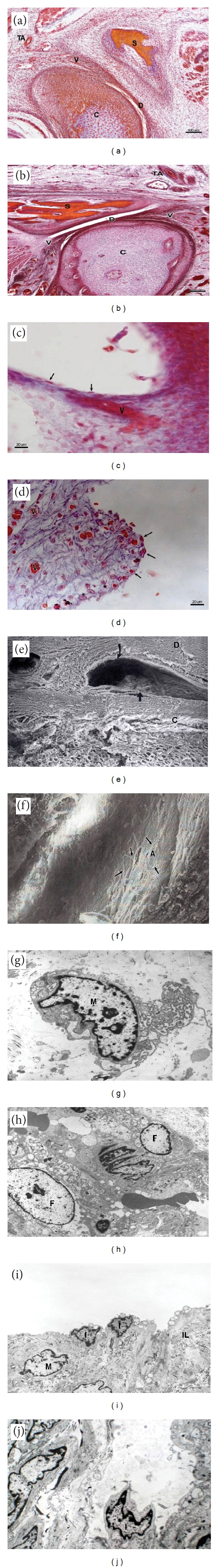
Objective. To characterize morphologically and ultrastructurally using light microscopy, the scanning electron microscopy and transmission electron microscopy the intima synovial of the temporomandibular joint (TMJ) of human fetuses between the 10th and the 38th week of development. Materials and Methods. The TMJ was dissected bilaterally in 37 human fetuses belonging to the Institute of Embryology of the University Complutense of Madrid and of the Federal University of São Paulo. Results. The outcome by light microscopy showed the morphology of the TMJ and that the formation of inferior joint cavity precedes the superior joint cavity and the presence of blood vessels in the synovial. Conclusion. By scanning and transmission electron microscopy we observed the presence of two well-defined cell types in the intima layer of synovial of the TMJ of human fetuses, macrophage-like type A cell and fibroblast-like type B cell, and the presence of the a third cell type, defined by the name of intermediate lining cell in the intima layer of the synovial.

 求助内容:
求助内容: 应助结果提醒方式:


