José Aderval Aragão, Luiza Neves de Santana Teles, Ana Bárbara de Jesus Chaves, Jéssica Cândida Oliveira Prado, Priscila Soares Pereira, João Gabriel Lima Dantas, Francisco Prado Reis
{"title":"The superior transverse scapular ligament in fetuses.","authors":"José Aderval Aragão, Luiza Neves de Santana Teles, Ana Bárbara de Jesus Chaves, Jéssica Cândida Oliveira Prado, Priscila Soares Pereira, João Gabriel Lima Dantas, Francisco Prado Reis","doi":"10.1155/2013/323194","DOIUrl":null,"url":null,"abstract":"<p><p>Introduction. The superior transverse scapular ligament (STSL) links the margins of the suprascapular notch and converts it into a foramen, through which, the suprascapular nerve and, on some rare occasions, the suprascapular vessels pass. This conversion often results from partial or complete ossification of the STSL and may produce compressive symptoms in the suprascapular nerve. Material and Method. Twenty shoulders from human fetuses were dissected without the aid of optical instruments and, using a digital pachymeter of precision 0.01 millimeters, length measurements and thickness measurements were made. The fetal age was from 21 to 33 weeks of gestation, with a mean of 27.6 ± 4.14 weeks. Results. There was no statistically significant difference in STSL length or any difference in the thicknesses at the medial and lateral extremities between the halves of the body (P ≥ 0.05). However, in the left half of the body, the medial extremity of the STSL was significantly thinner than the lateral extremity (P ≤ 0.05). Conclusion. Anatomical and morphometric details about the STSL were described in human fetuses. These findings, in fetuses, may encourage the pursuit of further studies to understand the morphofunctional role and meaning of this small ligament. </p>","PeriodicalId":89526,"journal":{"name":"Anatomy research international","volume":"2013 ","pages":"323194"},"PeriodicalIF":0.0000,"publicationDate":"2013-01-01","publicationTypes":"Journal Article","fieldsOfStudy":null,"isOpenAccess":false,"openAccessPdf":"https://sci-hub-pdf.com/10.1155/2013/323194","citationCount":"3","resultStr":null,"platform":"Semanticscholar","paperid":null,"PeriodicalName":"Anatomy research international","FirstCategoryId":"1085","ListUrlMain":"https://doi.org/10.1155/2013/323194","RegionNum":0,"RegionCategory":null,"ArticlePicture":[],"TitleCN":null,"AbstractTextCN":null,"PMCID":null,"EPubDate":"2013/12/18 0:00:00","PubModel":"Epub","JCR":"","JCRName":"","Score":null,"Total":0}
引用次数: 3
Abstract
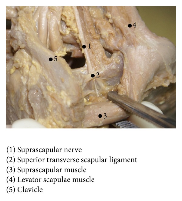
Introduction. The superior transverse scapular ligament (STSL) links the margins of the suprascapular notch and converts it into a foramen, through which, the suprascapular nerve and, on some rare occasions, the suprascapular vessels pass. This conversion often results from partial or complete ossification of the STSL and may produce compressive symptoms in the suprascapular nerve. Material and Method. Twenty shoulders from human fetuses were dissected without the aid of optical instruments and, using a digital pachymeter of precision 0.01 millimeters, length measurements and thickness measurements were made. The fetal age was from 21 to 33 weeks of gestation, with a mean of 27.6 ± 4.14 weeks. Results. There was no statistically significant difference in STSL length or any difference in the thicknesses at the medial and lateral extremities between the halves of the body (P ≥ 0.05). However, in the left half of the body, the medial extremity of the STSL was significantly thinner than the lateral extremity (P ≤ 0.05). Conclusion. Anatomical and morphometric details about the STSL were described in human fetuses. These findings, in fetuses, may encourage the pursuit of further studies to understand the morphofunctional role and meaning of this small ligament.

 求助内容:
求助内容: 应助结果提醒方式:


