Bahar Keles, Mutlu Duran, Yavuz Uyar, Ahmet Azimov, Abdullah Demirkan, Haci Hasan Esen
{"title":"Juvenile ossifying fibroma of the mandible: a case report.","authors":"Bahar Keles, Mutlu Duran, Yavuz Uyar, Ahmet Azimov, Abdullah Demirkan, Haci Hasan Esen","doi":"10.5037/jomr.2010.1205","DOIUrl":null,"url":null,"abstract":"<p><strong>Background: </strong>Fibro-osseous lesions of the jaws, including juvenile ossifying fibroma, pose diagnostic and therapeutic difficulties due to their clinical, radiological and histological variability. The aim of this study was to report the outcome of a 9 years old girl with diagnosed juvenile ossifying fibroma treatment.</p><p><strong>Methods: </strong>A 9 years old girl presented with a 6 x 8 cm sized hard fixed tumour on right ramus and corpus of the mandible. On the radiological examination tumour showed an irregular but well bordered, unilocular and expansive lesion on the right corpus and ramus of the mandible. There was no teeth displacement or teeth root resorbtion. Microscopically, the tumour had trabeculae, fibrillary osteoid and woven bone. After the clinical, radiological (panoramic radiography, computed tomography and magnetic resonance imaging) and histologic analysis it was diagnosed juvenile ossifying fibroma. In the history of the patient there has been an acute lymphocytic leukaemia in the remission for 3 years.</p><p><strong>Results: </strong>Because of large size of mandibular tumour, resultant expansion and destruction of mandibular cortex, the patient underwent right hemimandibulectomy using transmandibular approach. There was no recurrence or complications for two years follow-up.</p><p><strong>Conclusions: </strong>Although juvenile ossifying fibroma is an uncommon clinical entity, its aggressive local behaviour and high recurrence rate means that it is important to make an early diagnosis, apply the appropriate treatment and, especially, follow-up the patient over the long-term.</p>","PeriodicalId":53254,"journal":{"name":"eJournal of Oral Maxillofacial Research","volume":"1 2","pages":"e5"},"PeriodicalIF":1.0000,"publicationDate":"2010-07-01","publicationTypes":"Journal Article","fieldsOfStudy":null,"isOpenAccess":false,"openAccessPdf":"https://ftp.ncbi.nlm.nih.gov/pub/pmc/oa_pdf/d0/af/jomr-01-e5.PMC3886046.pdf","citationCount":"0","resultStr":null,"platform":"Semanticscholar","paperid":null,"PeriodicalName":"eJournal of Oral Maxillofacial Research","FirstCategoryId":"1085","ListUrlMain":"https://doi.org/10.5037/jomr.2010.1205","RegionNum":0,"RegionCategory":null,"ArticlePicture":[],"TitleCN":null,"AbstractTextCN":null,"PMCID":null,"EPubDate":"2010/1/1 0:00:00","PubModel":"eCollection","JCR":"Q3","JCRName":"DENTISTRY, ORAL SURGERY & MEDICINE","Score":null,"Total":0}
引用次数: 0
Abstract
Background: Fibro-osseous lesions of the jaws, including juvenile ossifying fibroma, pose diagnostic and therapeutic difficulties due to their clinical, radiological and histological variability. The aim of this study was to report the outcome of a 9 years old girl with diagnosed juvenile ossifying fibroma treatment.
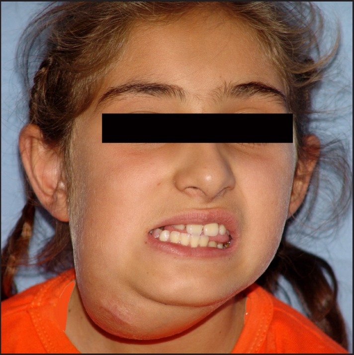
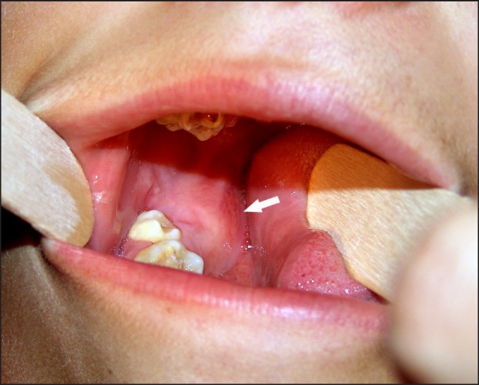
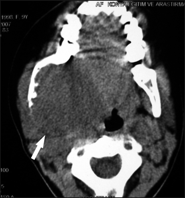
Methods: A 9 years old girl presented with a 6 x 8 cm sized hard fixed tumour on right ramus and corpus of the mandible. On the radiological examination tumour showed an irregular but well bordered, unilocular and expansive lesion on the right corpus and ramus of the mandible. There was no teeth displacement or teeth root resorbtion. Microscopically, the tumour had trabeculae, fibrillary osteoid and woven bone. After the clinical, radiological (panoramic radiography, computed tomography and magnetic resonance imaging) and histologic analysis it was diagnosed juvenile ossifying fibroma. In the history of the patient there has been an acute lymphocytic leukaemia in the remission for 3 years.
Results: Because of large size of mandibular tumour, resultant expansion and destruction of mandibular cortex, the patient underwent right hemimandibulectomy using transmandibular approach. There was no recurrence or complications for two years follow-up.
Conclusions: Although juvenile ossifying fibroma is an uncommon clinical entity, its aggressive local behaviour and high recurrence rate means that it is important to make an early diagnosis, apply the appropriate treatment and, especially, follow-up the patient over the long-term.

 求助内容:
求助内容: 应助结果提醒方式:


