{"title":"Scanning Electron Microscopic Studies of the Pecten Oculi in the Quail (Coturnix coturnix japonica).","authors":"Aris F Pourlis","doi":"10.1155/2013/650601","DOIUrl":null,"url":null,"abstract":"<p><p>The main purpose of this study is to extend the microscopic investigations of the pecten oculi in the quail in order to add some information on the unresolved functional anatomy of this unique avian organ. The pecten oculi of the quail was studied by scanning electron microscopy. Eighteen- to-twenty two highly vascularised accordion-like folds were joined apically by a heavily pigmented bridge of tissue, which holds the pecten in a fanlike shape, widest at the base. The structure of the double layered limiting membrane was recorded. The presence of hyalocytes with macrophage-like appearance was illustrated. It is assumed that the pecten oculi of the quail resembles that of the chicken. Illustrated morphological features of this species may add information on the active physiological role of the pecten. But still, the functional significance of this organ is a matter of controversies. </p>","PeriodicalId":89526,"journal":{"name":"Anatomy research international","volume":"2013 ","pages":"650601"},"PeriodicalIF":0.0000,"publicationDate":"2013-01-01","publicationTypes":"Journal Article","fieldsOfStudy":null,"isOpenAccess":false,"openAccessPdf":"https://sci-hub-pdf.com/10.1155/2013/650601","citationCount":"21","resultStr":null,"platform":"Semanticscholar","paperid":null,"PeriodicalName":"Anatomy research international","FirstCategoryId":"1085","ListUrlMain":"https://doi.org/10.1155/2013/650601","RegionNum":0,"RegionCategory":null,"ArticlePicture":[],"TitleCN":null,"AbstractTextCN":null,"PMCID":null,"EPubDate":"2013/10/2 0:00:00","PubModel":"Epub","JCR":"","JCRName":"","Score":null,"Total":0}
引用次数: 21
Abstract
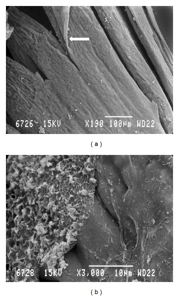
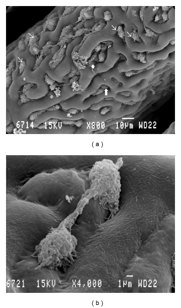
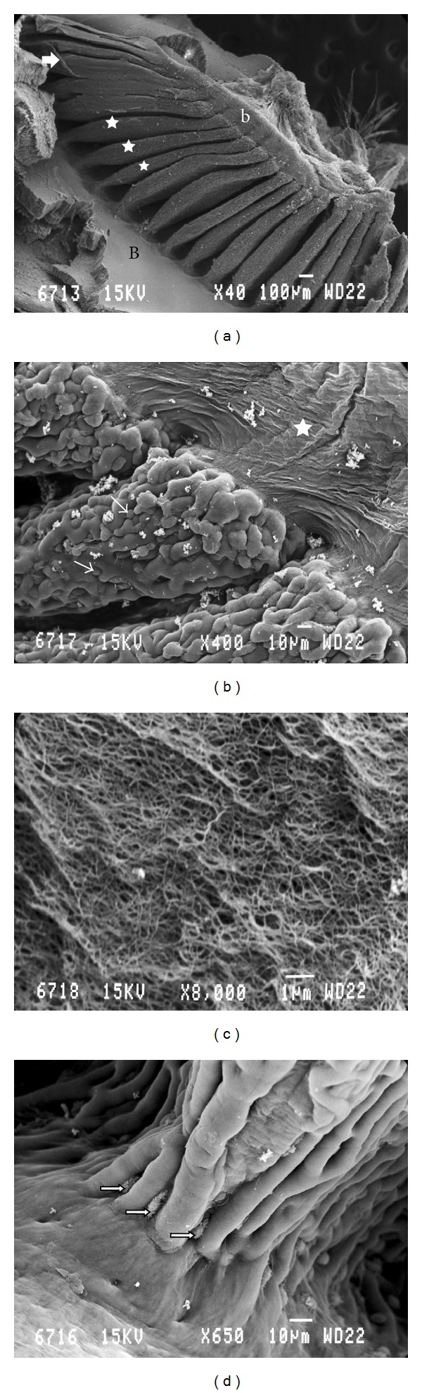
The main purpose of this study is to extend the microscopic investigations of the pecten oculi in the quail in order to add some information on the unresolved functional anatomy of this unique avian organ. The pecten oculi of the quail was studied by scanning electron microscopy. Eighteen- to-twenty two highly vascularised accordion-like folds were joined apically by a heavily pigmented bridge of tissue, which holds the pecten in a fanlike shape, widest at the base. The structure of the double layered limiting membrane was recorded. The presence of hyalocytes with macrophage-like appearance was illustrated. It is assumed that the pecten oculi of the quail resembles that of the chicken. Illustrated morphological features of this species may add information on the active physiological role of the pecten. But still, the functional significance of this organ is a matter of controversies.

 求助内容:
求助内容: 应助结果提醒方式:


