{"title":"A new cervical nerve root avulsion model using a posterior extra-vertebral approach in rats.","authors":"Takashi Noguchi, Souichi Ohta, Ryosuke Kakinoki, Yukitoshi Kaizawa, Shuichi Matsuda","doi":"10.1186/1749-7221-8-8","DOIUrl":null,"url":null,"abstract":"<p><strong>Background: </strong>The nerve root avulsion injury causes decrease of motor neurons in the spinal ventral horn. To investigate the motoneuron death after avulsion injury in rats, the intradural root avulsion procedure is usually used, although it is technically demanding and associated with a risk of unexpected spinal cord damage. We have developed a new cervical nerve root avulsion procedure in rats and investigated the validity of our procedure.</p><p><strong>Methods: </strong>Our procedure is using a posterior approach and pulling the C6 nerve root outside the vertebral foramen without intradural procedures. The lateral third of the lateral mass is needed to be resected before pulling the nerve root. The accomplishment of our procedure is judged by confirmation of the bifurcated stump of the avulsed nerve root and the leakage of the spinal fluid from vertebral foramen. At first, four Sprague-Dawley (SD) rats were used for the examination of C6 motor neuron distribution in the normal spinal cord. Then, 40 SD rats were divided into following four groups and the survival rate of motor neuron was examined. (A) an intradural avulsion group, (B) an intradural rhizotomy group, (C) our extravertebral avulsion group, and (D) an extravertebral rupture group. Another 26 SD rats were used for the examination of histomorphorogic changes in the spinal cord after our extra-vertebral avulsion procedure.</p><p><strong>Results: </strong>At 28 days after injury, the percentage of surviving motor neurons in groups A (39.0 ± 2.1%) and C (47.5 ± 7.1%) were significantly lower than those in groups B (77.1 ± 12.3%) and D (98.9 ± 9.9%). Compared with other groups, our procedure was easier and associated with less unexpected spinal cord damage. Although the length of the distal stump of the extravertebrally avulsed ventral rootlets was varied between 1.5 and 3.2 mm, this difference did not affect motoneuron death. The extravertebral avulsion injury showed intraspinal bleeding along the motoneuron axons, glial reaction and macrophage infiltration in the lesioned side of the ventral horn.</p><p><strong>Conclusions: </strong>Our extravertebral avulsion procedure is simple and reproducible. It would become a useful tool for the study of cervical nerve root avulsion injury.</p>","PeriodicalId":15280,"journal":{"name":"Journal of Brachial Plexus and Peripheral Nerve Injury","volume":"8 1","pages":"8"},"PeriodicalIF":1.1000,"publicationDate":"2013-09-11","publicationTypes":"Journal Article","fieldsOfStudy":null,"isOpenAccess":false,"openAccessPdf":"https://sci-hub-pdf.com/10.1186/1749-7221-8-8","citationCount":"11","resultStr":null,"platform":"Semanticscholar","paperid":null,"PeriodicalName":"Journal of Brachial Plexus and Peripheral Nerve Injury","FirstCategoryId":"1085","ListUrlMain":"https://doi.org/10.1186/1749-7221-8-8","RegionNum":0,"RegionCategory":null,"ArticlePicture":[],"TitleCN":null,"AbstractTextCN":null,"PMCID":null,"EPubDate":"","PubModel":"","JCR":"Q4","JCRName":"CLINICAL NEUROLOGY","Score":null,"Total":0}
引用次数: 11
Abstract
Background: The nerve root avulsion injury causes decrease of motor neurons in the spinal ventral horn. To investigate the motoneuron death after avulsion injury in rats, the intradural root avulsion procedure is usually used, although it is technically demanding and associated with a risk of unexpected spinal cord damage. We have developed a new cervical nerve root avulsion procedure in rats and investigated the validity of our procedure.
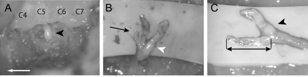
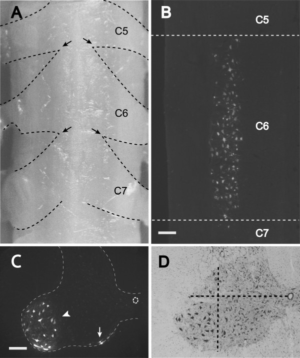
Methods: Our procedure is using a posterior approach and pulling the C6 nerve root outside the vertebral foramen without intradural procedures. The lateral third of the lateral mass is needed to be resected before pulling the nerve root. The accomplishment of our procedure is judged by confirmation of the bifurcated stump of the avulsed nerve root and the leakage of the spinal fluid from vertebral foramen. At first, four Sprague-Dawley (SD) rats were used for the examination of C6 motor neuron distribution in the normal spinal cord. Then, 40 SD rats were divided into following four groups and the survival rate of motor neuron was examined. (A) an intradural avulsion group, (B) an intradural rhizotomy group, (C) our extravertebral avulsion group, and (D) an extravertebral rupture group. Another 26 SD rats were used for the examination of histomorphorogic changes in the spinal cord after our extra-vertebral avulsion procedure.
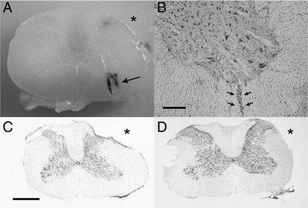
Results: At 28 days after injury, the percentage of surviving motor neurons in groups A (39.0 ± 2.1%) and C (47.5 ± 7.1%) were significantly lower than those in groups B (77.1 ± 12.3%) and D (98.9 ± 9.9%). Compared with other groups, our procedure was easier and associated with less unexpected spinal cord damage. Although the length of the distal stump of the extravertebrally avulsed ventral rootlets was varied between 1.5 and 3.2 mm, this difference did not affect motoneuron death. The extravertebral avulsion injury showed intraspinal bleeding along the motoneuron axons, glial reaction and macrophage infiltration in the lesioned side of the ventral horn.
Conclusions: Our extravertebral avulsion procedure is simple and reproducible. It would become a useful tool for the study of cervical nerve root avulsion injury.
期刊介绍:
JBPPNI is an open access, peer-reviewed online journal that will encompass all aspects of basic and clinical research findings, in the area of brachial plexus and peripheral nerve injury. Injury in this context refers to congenital, inflammatory, traumatic, degenerative and neoplastic processes, including neurofibromatosis. Papers on diagnostic and imaging aspects of the peripheral nervous system are welcomed as well. The peripheral nervous system is unique in its complexity and scope of influence. There are areas of interest in the anatomy, physiology, metabolism, phylogeny, and limb growth tropism of peripheral nerves.

 求助内容:
求助内容: 应助结果提醒方式:


