{"title":"Uterine cervical melanoma presenting with rapid progression detected by PET/CT.","authors":"Ya-Ju Tsai, Pei-Wei Shueng, Sheng-Chien Chan, Wen-Yu Chuang, Yu-Chien Shiau, Chung-Huei Hsu","doi":"10.1258/arsr.2012.120026","DOIUrl":null,"url":null,"abstract":"<p><p>Malignant melanoma of the uterine cervix is a rare extracutaneous melanoma which develops aggressively and is associated with a bleak prognosis. To our knowledge, no prior published reports have discussed the role of 18F-FDG positron emission tomography/computed tomography (PET/CT) in managing this disease. Our case study involved a 66-year-old woman with a malignant melanoma of the uterine cervix. The patient received PET/CT that identified metastases and lesions which had not been detected from her MRI. Serial PET/CT elucidated that the disease was initially limited to the pelvis, but then metastasized to the abdominal para-aortic lymph nodes, followed by extensive metastases to the brain, lungs, breast, supraclavicular, neck, and other abdominal lymph nodes, as observed at 6-month follow-up. PET/CT was used to complement conventional anatomic imaging modalities, and provided a novel modality for whole body screening. Visualization of the metabolic activity of indeterminate lesions may help in staging, re-staging, treatment planning, and prognostic prediction for patients with this rare disease. </p>","PeriodicalId":30445,"journal":{"name":"Acta Radiologica Short Reports","volume":"1 4","pages":""},"PeriodicalIF":0.0000,"publicationDate":"2012-05-17","publicationTypes":"Journal Article","fieldsOfStudy":null,"isOpenAccess":false,"openAccessPdf":"https://sci-hub-pdf.com/10.1258/arsr.2012.120026","citationCount":"5","resultStr":null,"platform":"Semanticscholar","paperid":null,"PeriodicalName":"Acta Radiologica Short Reports","FirstCategoryId":"1085","ListUrlMain":"https://doi.org/10.1258/arsr.2012.120026","RegionNum":0,"RegionCategory":null,"ArticlePicture":[],"TitleCN":null,"AbstractTextCN":null,"PMCID":null,"EPubDate":"2012/1/1 0:00:00","PubModel":"eCollection","JCR":"","JCRName":"","Score":null,"Total":0}
引用次数: 5
Abstract
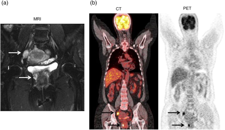
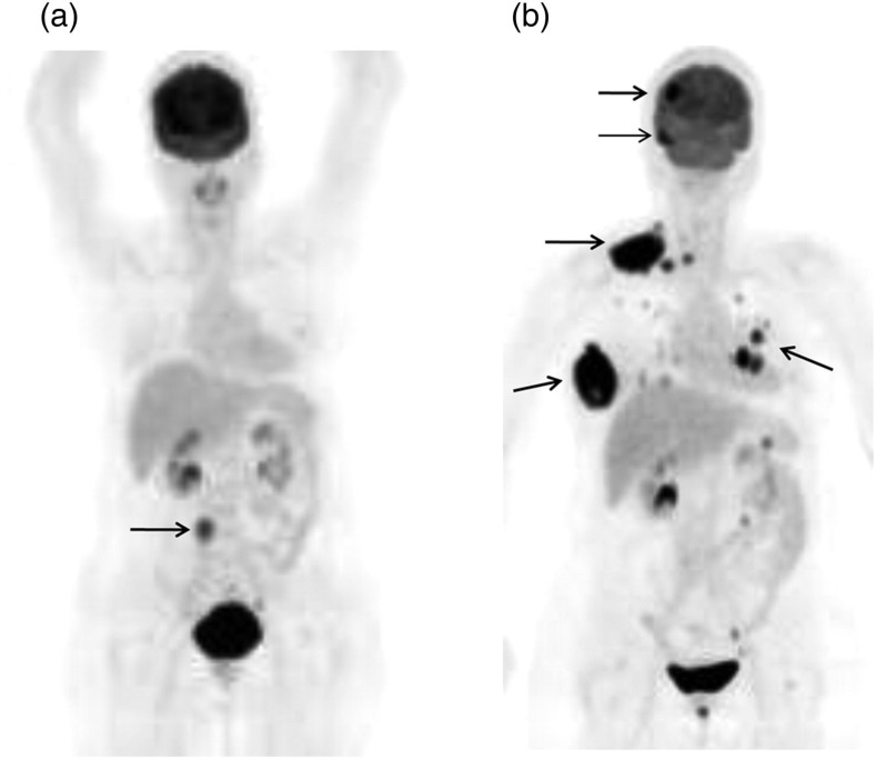
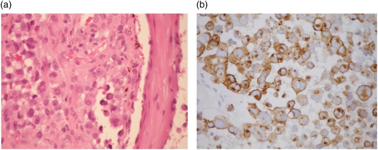
Malignant melanoma of the uterine cervix is a rare extracutaneous melanoma which develops aggressively and is associated with a bleak prognosis. To our knowledge, no prior published reports have discussed the role of 18F-FDG positron emission tomography/computed tomography (PET/CT) in managing this disease. Our case study involved a 66-year-old woman with a malignant melanoma of the uterine cervix. The patient received PET/CT that identified metastases and lesions which had not been detected from her MRI. Serial PET/CT elucidated that the disease was initially limited to the pelvis, but then metastasized to the abdominal para-aortic lymph nodes, followed by extensive metastases to the brain, lungs, breast, supraclavicular, neck, and other abdominal lymph nodes, as observed at 6-month follow-up. PET/CT was used to complement conventional anatomic imaging modalities, and provided a novel modality for whole body screening. Visualization of the metabolic activity of indeterminate lesions may help in staging, re-staging, treatment planning, and prognostic prediction for patients with this rare disease.
期刊介绍:
Under the editorial leadership of Professor Arnulf Skjennald MD and a distinguished editorial board, Acta Radiologica Open, formerly Acta Radiologica Short Reports, aims for the prompt publication of original case reports, short reports, review articles, pictorial reviews, research articles on diagnostic and interventional radiology, clinical radiology, experimental investigations in animals, and all other research related to imaging procedures. Acta Radiologica Open provides a complete update on all radiological specialties and technical utilities, as well as physiology and physics related to imaging, including ultrasonography, computed tomography, radionuclide and magnetic resonance imaging. Acta Radiologica Open publishes articles on diagnostic and interventional procedures in radiology based on all medical imaging techniques, as well as works in physiology and physics when related to radiology. The journal is an online-only, peer reviewed, open access journal.

 求助内容:
求助内容: 应助结果提醒方式:


