K Natsis, G Paraskevas, N Anastasopoulos, T Papamitsou, A Sioga
{"title":"Meniscofibular ligament: morphology and functional significance of a relatively unknown anatomical structure.","authors":"K Natsis, G Paraskevas, N Anastasopoulos, T Papamitsou, A Sioga","doi":"10.1155/2012/214784","DOIUrl":null,"url":null,"abstract":"<p><p>Purpose. A relatively unknown ligamentous structure of the posterolateral corner of the knee joint, the so-called meniscofibular ligament (MFL), was investigated as regards its macroscopic morphology, its histological features, and its reaction to knee movements. Material and Methods. MFL was exposed on 21 fresh-frozen unpaired knee joints. Its microscopic morphology was examined utilizing for comparison the fibular collateral and the popliteofibular ligament. Results. MFL was encountered in 100% of the specimens as a thin striplike fibrous band extending between the lower border of the lateral meniscus and the head of the fibula. MFL was tense during knee extension and external rotation of the tibia, whereas its histological features were similar to those of fibular collateral and popliteofibular ligament. Discussion. Its precise histological nature is studied as well as its tension alterations during knee movements. The potential functional significance of the MFL with respect to its role in avoidance of lateral meniscus and lateral coronary ligament tears is discussed. Conclusions. MFL presumably provides an additional protection to the lateral meniscus during the last stages of knee extension, as well as to the lateral coronary ligament reducing the possibility of a potential rupture.</p>","PeriodicalId":89526,"journal":{"name":"Anatomy research international","volume":"2012 ","pages":"214784"},"PeriodicalIF":0.0000,"publicationDate":"2012-01-01","publicationTypes":"Journal Article","fieldsOfStudy":null,"isOpenAccess":false,"openAccessPdf":"https://sci-hub-pdf.com/10.1155/2012/214784","citationCount":"19","resultStr":null,"platform":"Semanticscholar","paperid":null,"PeriodicalName":"Anatomy research international","FirstCategoryId":"1085","ListUrlMain":"https://doi.org/10.1155/2012/214784","RegionNum":0,"RegionCategory":null,"ArticlePicture":[],"TitleCN":null,"AbstractTextCN":null,"PMCID":null,"EPubDate":"2012/6/28 0:00:00","PubModel":"Epub","JCR":"","JCRName":"","Score":null,"Total":0}
引用次数: 19
Abstract
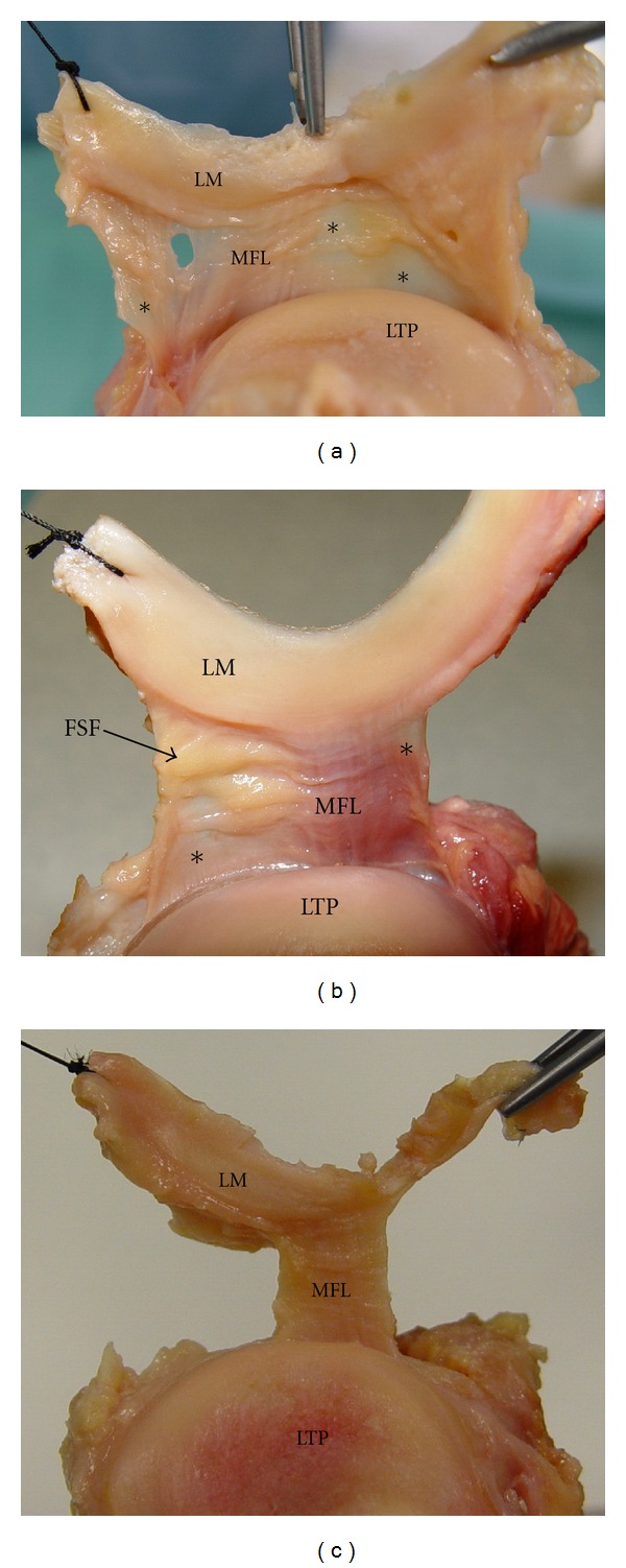
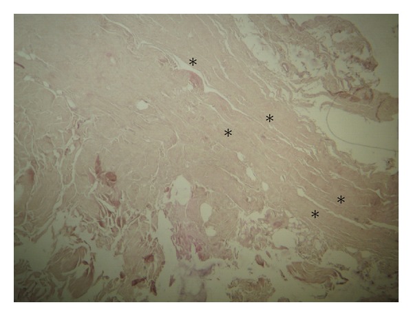
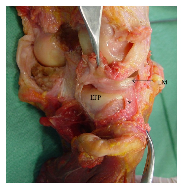
Purpose. A relatively unknown ligamentous structure of the posterolateral corner of the knee joint, the so-called meniscofibular ligament (MFL), was investigated as regards its macroscopic morphology, its histological features, and its reaction to knee movements. Material and Methods. MFL was exposed on 21 fresh-frozen unpaired knee joints. Its microscopic morphology was examined utilizing for comparison the fibular collateral and the popliteofibular ligament. Results. MFL was encountered in 100% of the specimens as a thin striplike fibrous band extending between the lower border of the lateral meniscus and the head of the fibula. MFL was tense during knee extension and external rotation of the tibia, whereas its histological features were similar to those of fibular collateral and popliteofibular ligament. Discussion. Its precise histological nature is studied as well as its tension alterations during knee movements. The potential functional significance of the MFL with respect to its role in avoidance of lateral meniscus and lateral coronary ligament tears is discussed. Conclusions. MFL presumably provides an additional protection to the lateral meniscus during the last stages of knee extension, as well as to the lateral coronary ligament reducing the possibility of a potential rupture.

 求助内容:
求助内容: 应助结果提醒方式:


