Imaging an adapted dentoalveolar complex.
引用次数: 15
Abstract
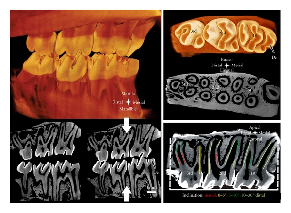
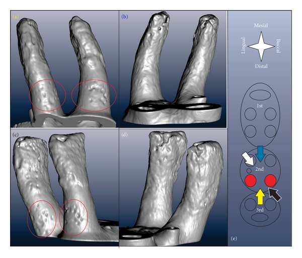
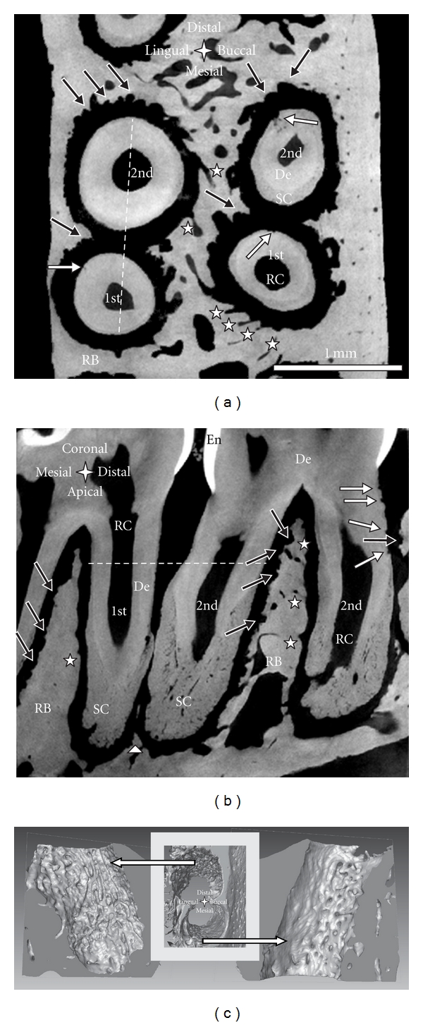
Adaptation of a rat dentoalveolar complex was illustrated using various imaging modalities. Micro-X-ray computed tomography for 3D modeling, combined with complementary techniques, including image processing, scanning electron microscopy, fluorochrome labeling, conventional histology (H&E, TRAP), and immunohistochemistry (RANKL, OPN) elucidated the dynamic nature of bone, the periodontal ligament-space, and cementum in the rat periodontium. Tomography and electron microscopy illustrated structural adaptation of calcified tissues at a higher resolution. Ongoing biomineralization was analyzed using fluorochrome labeling, and by evaluating attenuation profiles using virtual sections from 3D tomographies. Osteoclastic distribution as a function of anatomical location was illustrated by combining histology, immunohistochemistry, and tomography. While tomography and SEM provided past resorption-related events, future adaptive changes were deduced by identifying matrix biomolecules using immunohistochemistry. Thus, a dynamic picture of the dentoalveolar complex in rats was illustrated.
适应牙槽复合体成像。
适应大鼠牙槽复合体是用各种成像方式说明。用于三维建模的微x射线计算机断层扫描,结合互补技术,包括图像处理、扫描电子显微镜、荧光标记、常规组织学(H&E、TRAP)和免疫组织化学(RANKL、OPN),阐明了大鼠牙周组织中骨、牙周韧带间隙和牙骨质的动态特性。断层扫描和电子显微镜以更高的分辨率显示钙化组织的结构适应。正在进行的生物矿化分析使用荧光标记,并通过评估衰减剖面从三维断层扫描的虚拟切片。通过结合组织学、免疫组织化学和断层扫描来说明破骨细胞分布作为解剖位置的功能。虽然断层扫描和扫描电镜提供了过去的吸收相关事件,但未来的适应性变化是通过免疫组织化学鉴定基质生物分子来推断的。因此,在大鼠牙槽嵴复合体的动态图片被说明。
本文章由计算机程序翻译,如有差异,请以英文原文为准。
求助全文
约1分钟内获得全文
求助全文

 求助内容:
求助内容: 应助结果提醒方式:


