{"title":"Optimizing automated characterization of liver fibrosis histological images by investigating color spaces at different resolutions.","authors":"Doaa Mahmoud-Ghoneim","doi":"10.1186/1742-4682-8-25","DOIUrl":null,"url":null,"abstract":"<p><p>Texture analysis (TA) of histological images has recently received attention as an automated method of characterizing liver fibrosis. The colored staining methods used to identify different tissue components reveal various patterns that contribute in different ways to the digital texture of the image. A histological digital image can be represented with various color spaces. The approximation processes of pixel values that are carried out while converting between different color spaces can affect image texture and subsequently could influence the performance of TA. Conventional TA is carried out on grey scale images, which are a luminance approximation to the original RGB (Red, Green, and Blue) space. Currently, grey scale is considered sufficient for characterization of fibrosis but this may not be the case for sophisticated assessment of fibrosis or when resolution conditions vary. This paper investigates the accuracy of TA results on three color spaces, conventional grey scale, RGB, and Hue-Saturation-Intensity (HSI), at different resolutions. The results demonstrate that RGB is the most accurate in texture classification of liver images, producing better results, most notably at low resolution. Furthermore, the green channel, which is dominated by collagen fiber deposition, appears to provide most of the features for characterizing fibrosis images. The HSI space demonstrated a high percentage error for the majority of texture methods at all resolutions, suggesting that this space is insufficient for fibrosis characterization. The grey scale space produced good results at high resolution; however, errors increased as resolution decreased.</p>","PeriodicalId":51195,"journal":{"name":"Theoretical Biology and Medical Modelling","volume":" ","pages":"25"},"PeriodicalIF":0.0000,"publicationDate":"2011-07-14","publicationTypes":"Journal Article","fieldsOfStudy":null,"isOpenAccess":false,"openAccessPdf":"https://sci-hub-pdf.com/10.1186/1742-4682-8-25","citationCount":"14","resultStr":null,"platform":"Semanticscholar","paperid":null,"PeriodicalName":"Theoretical Biology and Medical Modelling","FirstCategoryId":"1085","ListUrlMain":"https://doi.org/10.1186/1742-4682-8-25","RegionNum":0,"RegionCategory":null,"ArticlePicture":[],"TitleCN":null,"AbstractTextCN":null,"PMCID":null,"EPubDate":"","PubModel":"","JCR":"Q1","JCRName":"Mathematics","Score":null,"Total":0}
引用次数: 14
Abstract
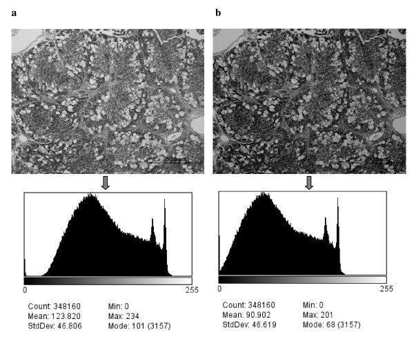
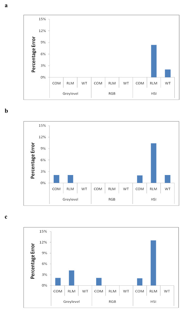
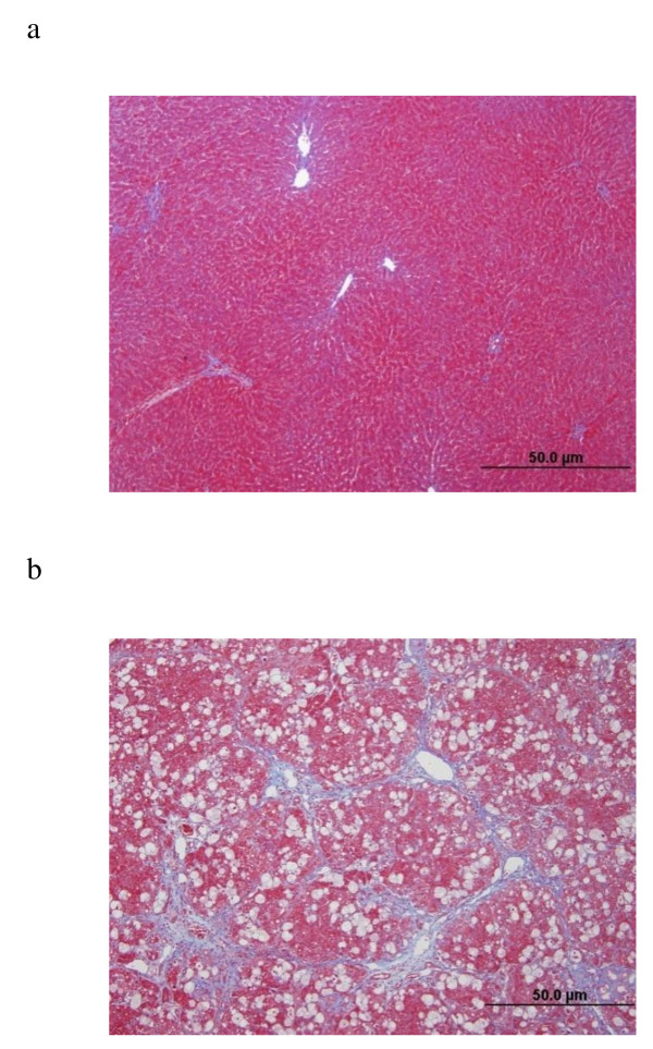
Texture analysis (TA) of histological images has recently received attention as an automated method of characterizing liver fibrosis. The colored staining methods used to identify different tissue components reveal various patterns that contribute in different ways to the digital texture of the image. A histological digital image can be represented with various color spaces. The approximation processes of pixel values that are carried out while converting between different color spaces can affect image texture and subsequently could influence the performance of TA. Conventional TA is carried out on grey scale images, which are a luminance approximation to the original RGB (Red, Green, and Blue) space. Currently, grey scale is considered sufficient for characterization of fibrosis but this may not be the case for sophisticated assessment of fibrosis or when resolution conditions vary. This paper investigates the accuracy of TA results on three color spaces, conventional grey scale, RGB, and Hue-Saturation-Intensity (HSI), at different resolutions. The results demonstrate that RGB is the most accurate in texture classification of liver images, producing better results, most notably at low resolution. Furthermore, the green channel, which is dominated by collagen fiber deposition, appears to provide most of the features for characterizing fibrosis images. The HSI space demonstrated a high percentage error for the majority of texture methods at all resolutions, suggesting that this space is insufficient for fibrosis characterization. The grey scale space produced good results at high resolution; however, errors increased as resolution decreased.
期刊介绍:
Theoretical Biology and Medical Modelling is an open access peer-reviewed journal adopting a broad definition of "biology" and focusing on theoretical ideas and models associated with developments in biology and medicine. Mathematicians, biologists and clinicians of various specialisms, philosophers and historians of science are all contributing to the emergence of novel concepts in an age of systems biology, bioinformatics and computer modelling. This is the field in which Theoretical Biology and Medical Modelling operates. We welcome submissions that are technically sound and offering either improved understanding in biology and medicine or progress in theory or method.
 求助内容:
求助内容: 应助结果提醒方式:


