{"title":"Adenomatoid tumor of testis.","authors":"Waqas Amin, Anil V Parwani","doi":"10.4137/cpath.s3091","DOIUrl":null,"url":null,"abstract":"<p><p>Adenomatoid tumors are responsible for 30% of all paratesticular masses. These are usually asymptomatic, slow growing masses. They are benign tumors comprising of cords and tubules of cuboidal to columnar cells with vacuolated cytoplasm and fibrous stroma. They are considered to be of mesothelial origin supported by histochemical studies and genetic analysis of Wilms tumor 1 gene expression. Excision biopsy is both diagnostic and therapeutic procedure. The main clinical consideration is accurate diagnosis preventing unnecessary orchiectomy. Diagnostic studies include serum tumor markers (negative alpha fetoprotein, beta HCG, LDH) ultrasonography (hypoechoic and homogenous appearance) and frozen section.</p>","PeriodicalId":89118,"journal":{"name":"Clinical medicine. Pathology","volume":"2 ","pages":"17-22"},"PeriodicalIF":0.0000,"publicationDate":"2009-09-09","publicationTypes":"Journal Article","fieldsOfStudy":null,"isOpenAccess":false,"openAccessPdf":"https://sci-hub-pdf.com/10.4137/cpath.s3091","citationCount":"33","resultStr":null,"platform":"Semanticscholar","paperid":null,"PeriodicalName":"Clinical medicine. Pathology","FirstCategoryId":"1085","ListUrlMain":"https://doi.org/10.4137/cpath.s3091","RegionNum":0,"RegionCategory":null,"ArticlePicture":[],"TitleCN":null,"AbstractTextCN":null,"PMCID":null,"EPubDate":"","PubModel":"","JCR":"","JCRName":"","Score":null,"Total":0}
引用次数: 33
Abstract
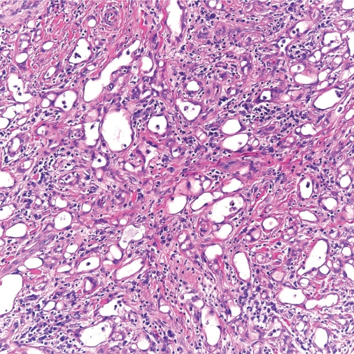
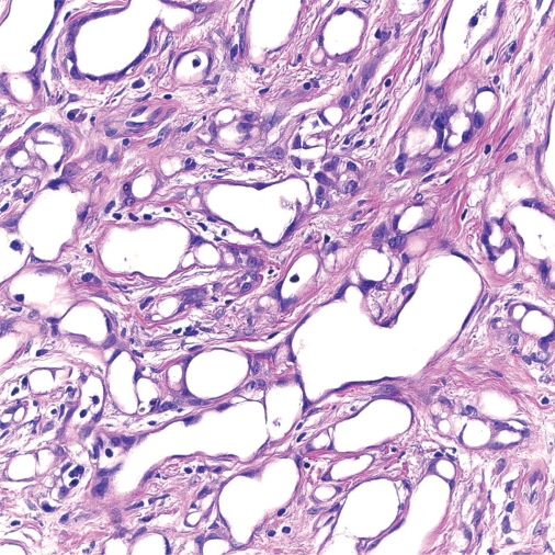
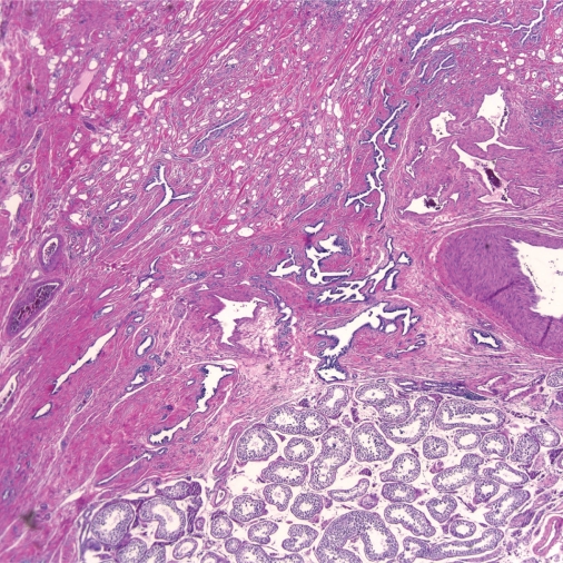
Adenomatoid tumors are responsible for 30% of all paratesticular masses. These are usually asymptomatic, slow growing masses. They are benign tumors comprising of cords and tubules of cuboidal to columnar cells with vacuolated cytoplasm and fibrous stroma. They are considered to be of mesothelial origin supported by histochemical studies and genetic analysis of Wilms tumor 1 gene expression. Excision biopsy is both diagnostic and therapeutic procedure. The main clinical consideration is accurate diagnosis preventing unnecessary orchiectomy. Diagnostic studies include serum tumor markers (negative alpha fetoprotein, beta HCG, LDH) ultrasonography (hypoechoic and homogenous appearance) and frozen section.

 求助内容:
求助内容: 应助结果提醒方式:


