Mu-Gen Liu, Hong Li, Xuming Xu, Colin J Barnstable, Samuel Shao-Min Zhang
{"title":"Comparison of gene expression during in vivo and in vitro postnatal retina development.","authors":"Mu-Gen Liu, Hong Li, Xuming Xu, Colin J Barnstable, Samuel Shao-Min Zhang","doi":"10.1007/s12177-008-9009-z","DOIUrl":null,"url":null,"abstract":"<p><strong>Unlabelled: </strong>Retina explants are widely used as a model of neural development. To define the molecular basis of differences between the development of retina in vivo and in vitro during the early postnatal period, we carried out a series of microarray comparisons using mouse retinas. About 75% of 8,880 expressed genes from retina explants kept the same expression volume and pattern as the retina in vivo. Fewer than 6% of the total gene population was changed at two consecutive time points, and only about 1% genes showed more than a threefold change at any time point studied. Functional Gene Ontology (GO) mapping for both changed and unchanged genes showed similar distribution patterns, except that more genes were changed in the GO clusters of response to stimuli and carbohydrate metabolism. Three distinct expression patterns of genes preferentially expressed in rod photoreceptors were observed in the retina explants. Some genes showed a lag in increased expression, some showed no change, and some continued to have a reduced level of expression. An early downregulation of cyclin D1 in the explanted retina might explain the reduction in numbers of precursors in explanted retina and suggests that external factors are required for maintenance of cyclin D1. The global view of gene profiles presented in this study will help define the molecular changes in retina explants over time and will provide criteria to define future changes that improve this model system.</p><p><strong>Electronic supplementary material: </strong>The online version of this article (doi:10.1007/s12177-008-9009-z) contains supplementary material, which is available to authorized users.</p>","PeriodicalId":73873,"journal":{"name":"Journal of ocular biology, diseases, and informatics","volume":"1 2-4","pages":"59-72"},"PeriodicalIF":0.0000,"publicationDate":"2008-12-01","publicationTypes":"Journal Article","fieldsOfStudy":null,"isOpenAccess":false,"openAccessPdf":"https://sci-hub-pdf.com/10.1007/s12177-008-9009-z","citationCount":"13","resultStr":null,"platform":"Semanticscholar","paperid":null,"PeriodicalName":"Journal of ocular biology, diseases, and informatics","FirstCategoryId":"1085","ListUrlMain":"https://doi.org/10.1007/s12177-008-9009-z","RegionNum":0,"RegionCategory":null,"ArticlePicture":[],"TitleCN":null,"AbstractTextCN":null,"PMCID":null,"EPubDate":"2008/7/11 0:00:00","PubModel":"Epub","JCR":"","JCRName":"","Score":null,"Total":0}
引用次数: 13
Abstract
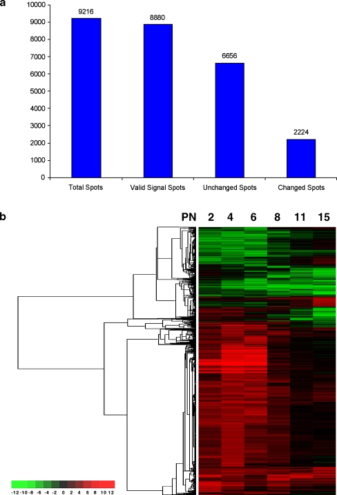
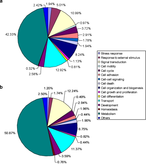
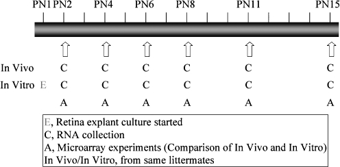
Unlabelled: Retina explants are widely used as a model of neural development. To define the molecular basis of differences between the development of retina in vivo and in vitro during the early postnatal period, we carried out a series of microarray comparisons using mouse retinas. About 75% of 8,880 expressed genes from retina explants kept the same expression volume and pattern as the retina in vivo. Fewer than 6% of the total gene population was changed at two consecutive time points, and only about 1% genes showed more than a threefold change at any time point studied. Functional Gene Ontology (GO) mapping for both changed and unchanged genes showed similar distribution patterns, except that more genes were changed in the GO clusters of response to stimuli and carbohydrate metabolism. Three distinct expression patterns of genes preferentially expressed in rod photoreceptors were observed in the retina explants. Some genes showed a lag in increased expression, some showed no change, and some continued to have a reduced level of expression. An early downregulation of cyclin D1 in the explanted retina might explain the reduction in numbers of precursors in explanted retina and suggests that external factors are required for maintenance of cyclin D1. The global view of gene profiles presented in this study will help define the molecular changes in retina explants over time and will provide criteria to define future changes that improve this model system.
Electronic supplementary material: The online version of this article (doi:10.1007/s12177-008-9009-z) contains supplementary material, which is available to authorized users.

 求助内容:
求助内容: 应助结果提醒方式:


