{"title":"Biocompatibility of subretinal parylene-based Ti/Pt microelectrode array in rabbit for further artificial vision studies.","authors":"Weihong Yu, Xuqian Wang, Chan Zhao, Zhikun Yang, Rongping Dai, Fangtian Dong","doi":"10.1007/s12177-009-9018-6","DOIUrl":null,"url":null,"abstract":"<p><p>To evaluate the biocompatibility of subretinal implanted parylene-based Ti/Pt microelectrode arrays (MEA). Eyes were enucleated 3 months after MEAs were implanted into the subretinal space of rabbits. Morphological changes of the retinas were investigated by H&E staining. Immunohistochemical staining for glial fibrillary acidic protein and opsin were performed to evaluate changes in Muller cells and photoreceptors in the retinas. Retina tissue around the array remained intact. Photoreceptor degeneration and glial cell activation were observed in the retina overlaying the MEA implant. However, the cells in the inner retinal layers were preserved. Photoreceptor degeneration and glial cell activation at the MEA-retina interface are expected to be a normal reaction to implantation. Material used in this experiment has good biocompatibility within the subretinal environment and is expected to be promising in the further retinal prosthesis studies.</p>","PeriodicalId":73873,"journal":{"name":"Journal of ocular biology, diseases, and informatics","volume":"2 1","pages":"33-6"},"PeriodicalIF":0.0000,"publicationDate":"2009-03-27","publicationTypes":"Journal Article","fieldsOfStudy":null,"isOpenAccess":false,"openAccessPdf":"https://sci-hub-pdf.com/10.1007/s12177-009-9018-6","citationCount":"12","resultStr":null,"platform":"Semanticscholar","paperid":null,"PeriodicalName":"Journal of ocular biology, diseases, and informatics","FirstCategoryId":"1085","ListUrlMain":"https://doi.org/10.1007/s12177-009-9018-6","RegionNum":0,"RegionCategory":null,"ArticlePicture":[],"TitleCN":null,"AbstractTextCN":null,"PMCID":null,"EPubDate":"","PubModel":"","JCR":"","JCRName":"","Score":null,"Total":0}
引用次数: 12
Abstract
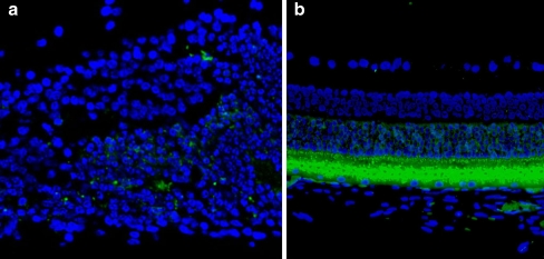
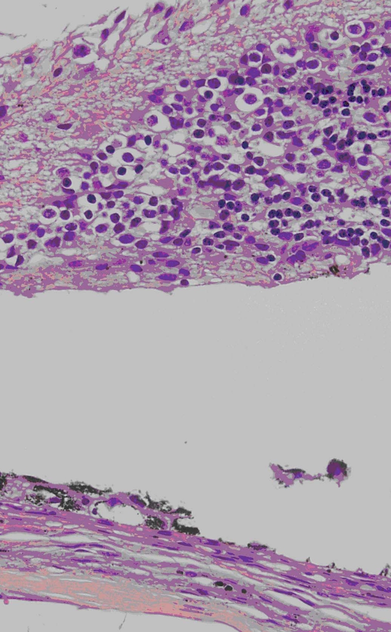
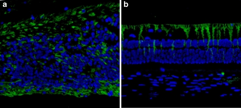
To evaluate the biocompatibility of subretinal implanted parylene-based Ti/Pt microelectrode arrays (MEA). Eyes were enucleated 3 months after MEAs were implanted into the subretinal space of rabbits. Morphological changes of the retinas were investigated by H&E staining. Immunohistochemical staining for glial fibrillary acidic protein and opsin were performed to evaluate changes in Muller cells and photoreceptors in the retinas. Retina tissue around the array remained intact. Photoreceptor degeneration and glial cell activation were observed in the retina overlaying the MEA implant. However, the cells in the inner retinal layers were preserved. Photoreceptor degeneration and glial cell activation at the MEA-retina interface are expected to be a normal reaction to implantation. Material used in this experiment has good biocompatibility within the subretinal environment and is expected to be promising in the further retinal prosthesis studies.

 求助内容:
求助内容: 应助结果提醒方式:


