Structural Alterations of Epididymal Epithelial Cells in Cathepsin A—Deficient Mice Affect the Blood-Epididymal Barrier and Lead to Altered Sperm Motility
Louis Hermo, Nadine Korah, Mary Gregory, Lauren Ye Liu, Daniel G. Cyr, Alessandra D'Azzo, Charles E. Smith
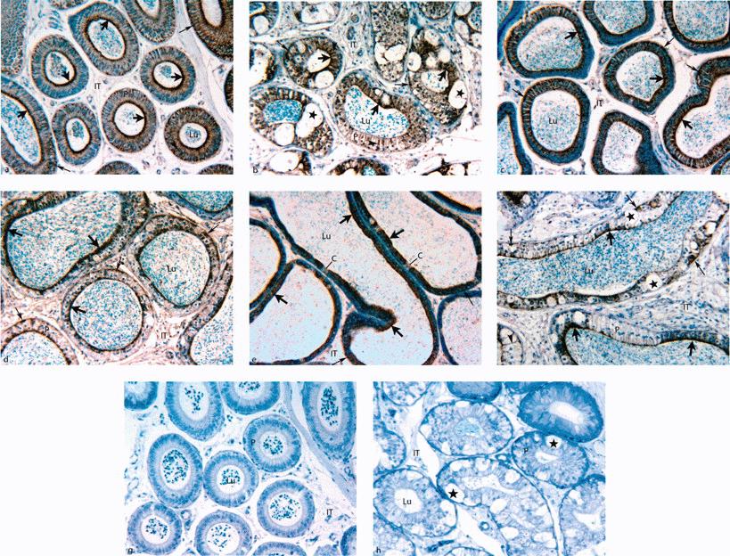
{"title":"Structural Alterations of Epididymal Epithelial Cells in Cathepsin A—Deficient Mice Affect the Blood-Epididymal Barrier and Lead to Altered Sperm Motility","authors":"Louis Hermo, Nadine Korah, Mary Gregory, Lauren Ye Liu, Daniel G. Cyr, Alessandra D'Azzo, Charles E. Smith","doi":"10.2164/jandrol.107.002980","DOIUrl":null,"url":null,"abstract":"<p><b>ABSTRACT: </b> Past studies have shown that the epithelial lining of the epididymis in adult mice deficient in protective protein cathepsin A (PPCA −/−) becomes swollen and vacuolated as a result of an accumulation of pale lysosomes, some very large, in addition to the presence of an abundance of macrophages infiltrating the intertubular spaces. The purpose of this study was to assess the integrity of the epididymal epithelial—blood barrier in these altered mice by characterizing the distribution of claudins (Cldns) and the leakiness of tight junctions to lanthanum nitrate. A second goal was to characterize sperm motility behavior in PPCA −/− mice using computer-assisted sperm analyses (CASA). The results indicated that lanthanum nitrate penetrated apical junctional complexes between adjacent epithelial cells and entered the epididymal lumen in PPCA −/− mice but not in control PPCA +/+ mice. Immunostaining for Cldns 1, 3, 8, and 10 revealed unique patterns of expression based on cell type and region specificity in PPCA +/+ mice, which were much different in PPCA –/– mice. PPCA –/– mice showed reduced intensities of immunoreactions, complete absence of immunoreactions, and appearance of atypical cytoplasmic immunoreactions. CASA indicated that sperm counts in the PPCA –/– mice were 70% reduced, and among other problems, there was a fourfold higher percentage of static sperm in PPCA –/– mice compared with controls. These results suggest that PPCA deficiency causes structural changes to the blood-epididymal barrier as evidenced by lanthanum nitrate and Cldns expression that affects the luminal environment of the epididymis, resulting in altered sperm motility.</p>","PeriodicalId":15029,"journal":{"name":"Journal of andrology","volume":"28 5","pages":"784-797"},"PeriodicalIF":0.0000,"publicationDate":"2013-01-02","publicationTypes":"Journal Article","fieldsOfStudy":null,"isOpenAccess":false,"openAccessPdf":"https://sci-hub-pdf.com/10.2164/jandrol.107.002980","citationCount":"26","resultStr":null,"platform":"Semanticscholar","paperid":null,"PeriodicalName":"Journal of andrology","FirstCategoryId":"1085","ListUrlMain":"https://onlinelibrary.wiley.com/doi/10.2164/jandrol.107.002980","RegionNum":0,"RegionCategory":null,"ArticlePicture":[],"TitleCN":null,"AbstractTextCN":null,"PMCID":null,"EPubDate":"","PubModel":"","JCR":"","JCRName":"","Score":null,"Total":0}
引用次数: 26
Abstract
ABSTRACT: Past studies have shown that the epithelial lining of the epididymis in adult mice deficient in protective protein cathepsin A (PPCA −/−) becomes swollen and vacuolated as a result of an accumulation of pale lysosomes, some very large, in addition to the presence of an abundance of macrophages infiltrating the intertubular spaces. The purpose of this study was to assess the integrity of the epididymal epithelial—blood barrier in these altered mice by characterizing the distribution of claudins (Cldns) and the leakiness of tight junctions to lanthanum nitrate. A second goal was to characterize sperm motility behavior in PPCA −/− mice using computer-assisted sperm analyses (CASA). The results indicated that lanthanum nitrate penetrated apical junctional complexes between adjacent epithelial cells and entered the epididymal lumen in PPCA −/− mice but not in control PPCA +/+ mice. Immunostaining for Cldns 1, 3, 8, and 10 revealed unique patterns of expression based on cell type and region specificity in PPCA +/+ mice, which were much different in PPCA –/– mice. PPCA –/– mice showed reduced intensities of immunoreactions, complete absence of immunoreactions, and appearance of atypical cytoplasmic immunoreactions. CASA indicated that sperm counts in the PPCA –/– mice were 70% reduced, and among other problems, there was a fourfold higher percentage of static sperm in PPCA –/– mice compared with controls. These results suggest that PPCA deficiency causes structural changes to the blood-epididymal barrier as evidenced by lanthanum nitrate and Cldns expression that affects the luminal environment of the epididymis, resulting in altered sperm motility.

 求助内容:
求助内容: 应助结果提醒方式:


