{"title":"Magnetic Resonance Imaging of Orbital Solitary Fibrous Tumors: Radiological-Pathological Correlation Analysis.","authors":"Ryuhei Masuno, Daisuke Yunaiyama, Yukiko Shishido-Hara, Daisuke Yoshimaru, Chifumi Maruyama, Yoichi Araki, Hiroshi Goto, Toshitaka Nagao, Kazuhiro Saito","doi":"10.5334/jbsr.2097","DOIUrl":null,"url":null,"abstract":"<p><strong>Background: </strong>Solitary fibrous tumors (SFTs) are rare and can be misdiagnosed because of their various radiological appearances.</p><p><strong>Purpose: </strong>To clarify the characteristic MRI findings of SFTs by analyzing their radiological-pathological correlation.</p><p><strong>Material and methods: </strong>Nine consecutive patients with SFT who underwent magnetic resonance imaging (MRI) prior to surgery were analyzed. Eight patients underwent contrast-enhanced MRI, and three underwent dynamic MRI. Radiological-pathological correlation analysis, co-occurrence matrix, run-length matrix, and histogram analysis were performed to assess the relationship between pathological findings T1- and T2-weighted images (T1-WI and T2-WI).</p><p><strong>Results: </strong>All nine lesions ranged in size from 20 to 36 mm. Seven lesions were located in the superior portion of the retrobulbar space found outside of the muscle cone, and two lesions in the inferior portion were located within it. No significant correlation was observed between the amount of collagenous tissue and the qualitative evaluation of the signal on T1-WI and T2-WI. Kurtosis on T2-WI was significantly correlated with the amount of collagenous tissue (<i>ρ</i> = -0.97, <i>p</i> < 0.0001) and endothelial cells (<i>ρ</i> = -0.49, <i>p</i> = 0.0479).</p><p><strong>Conclusion: </strong>Kurtosis in the histogram analysis on T2WI showed a strong correlation with the amount of collagenous tissue.</p>","PeriodicalId":56282,"journal":{"name":"Journal of the Belgian Society of Radiology","volume":"105 1","pages":"14"},"PeriodicalIF":1.3000,"publicationDate":"2021-03-16","publicationTypes":"Journal Article","fieldsOfStudy":null,"isOpenAccess":false,"openAccessPdf":"https://www.ncbi.nlm.nih.gov/pmc/articles/PMC7977021/pdf/","citationCount":"2","resultStr":null,"platform":"Semanticscholar","paperid":null,"PeriodicalName":"Journal of the Belgian Society of Radiology","FirstCategoryId":"3","ListUrlMain":"https://doi.org/10.5334/jbsr.2097","RegionNum":4,"RegionCategory":"医学","ArticlePicture":[],"TitleCN":null,"AbstractTextCN":null,"PMCID":null,"EPubDate":"","PubModel":"","JCR":"Q4","JCRName":"Medicine","Score":null,"Total":0}
引用次数: 2
Abstract
Background: Solitary fibrous tumors (SFTs) are rare and can be misdiagnosed because of their various radiological appearances.
Purpose: To clarify the characteristic MRI findings of SFTs by analyzing their radiological-pathological correlation.
Material and methods: Nine consecutive patients with SFT who underwent magnetic resonance imaging (MRI) prior to surgery were analyzed. Eight patients underwent contrast-enhanced MRI, and three underwent dynamic MRI. Radiological-pathological correlation analysis, co-occurrence matrix, run-length matrix, and histogram analysis were performed to assess the relationship between pathological findings T1- and T2-weighted images (T1-WI and T2-WI).
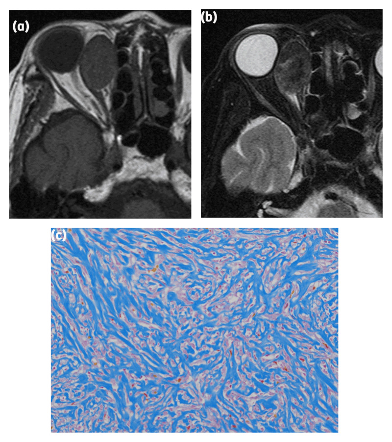
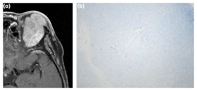
Results: All nine lesions ranged in size from 20 to 36 mm. Seven lesions were located in the superior portion of the retrobulbar space found outside of the muscle cone, and two lesions in the inferior portion were located within it. No significant correlation was observed between the amount of collagenous tissue and the qualitative evaluation of the signal on T1-WI and T2-WI. Kurtosis on T2-WI was significantly correlated with the amount of collagenous tissue (ρ = -0.97, p < 0.0001) and endothelial cells (ρ = -0.49, p = 0.0479).
Conclusion: Kurtosis in the histogram analysis on T2WI showed a strong correlation with the amount of collagenous tissue.
期刊介绍:
The purpose of the Journal of the Belgian Society of Radiology is the publication of articles dealing with diagnostic and interventional radiology, related imaging techniques, allied sciences, and continuing education.

 求助内容:
求助内容: 应助结果提醒方式:


