Natascha Schurz, Lydia Sariaslani, Patrick Altmann, Fritz Leutmezer, Christoph Mitsch, Berthold Pemp, Paulus Rommer, Tobias Zrzavy, Thomas Berger, Gabriel Bsteh
{"title":"Evaluation of Retinal Layer Thickness Parameters as Biomarkers in a Real-World Multiple Sclerosis Cohort.","authors":"Natascha Schurz, Lydia Sariaslani, Patrick Altmann, Fritz Leutmezer, Christoph Mitsch, Berthold Pemp, Paulus Rommer, Tobias Zrzavy, Thomas Berger, Gabriel Bsteh","doi":"10.2147/EB.S295610","DOIUrl":null,"url":null,"abstract":"<p><strong>Purpose: </strong>Retinal layer thickness parameters measured by optical coherence tomography (OCT) are emerging biomarkers of neuroaxonal degeneration and inflammation in multiple sclerosis (MS). We aimed to evaluate the value of retinal layer thickness for prediction of disability worsening and relapse in a real-world MS cohort.</p><p><strong>Patients and methods: </strong>For this longitudinal observational study, we included MS patients with spectral-domain OCT scans available and ≥1 year of clinical follow-up. The value of peripapillary retinal nerve fiber layer (pRNFL), macular ganglion-cell-and-inner-plexiform-layer (GCIPL) and inner nuclear layer (INL) thickness for prediction of disability worsening and relapse during the observation period was tested by multivariate models.</p><p><strong>Results: </strong>We analyzed 60 MS patients during a mean observation period of 2.9 years (SD 1.8). Lower baseline thickness of GCIPL (cut-off <77µm; HR 4.1, p=0.001) and pRNFL (cut-off ≤88µm; HR 3.1, p=0.019) were associated with an increased risk of disability worsening. Longitudinally, mean thinning rates were -0.8µm/year (SD 1.6) for GCIPL, -0.6µm/year (SD 3.5) for pRNFL. GCIPL thinning ≥1.0µm/year and pRNFL >1.5µm/year is associated with higher likelihood of disability worsening (HR 5.7, p=0.009 and HR 6.8, p=0.003, respectively). INL thickened in patients with relapse by a mean 0.9µm while thinning by 0.3µm in patients without relapse (p=0.04). In multivariate analyses, INL thickening was associated with an increased probability of relapse (OR 17.8, p=0.023).</p><p><strong>Conclusion: </strong>Cross-sectional and longitudinal measurement of GCIPL and pRNFL thinning is reliable as a biomarker of disability worsening in a real-world setting. Change of INL thickness is a promising marker of relapse, i.e. inflammatory activity.</p>","PeriodicalId":51844,"journal":{"name":"Eye and Brain","volume":"13 ","pages":"59-69"},"PeriodicalIF":2.4000,"publicationDate":"2021-03-12","publicationTypes":"Journal Article","fieldsOfStudy":null,"isOpenAccess":false,"openAccessPdf":"https://ftp.ncbi.nlm.nih.gov/pub/pmc/oa_pdf/76/89/eb-13-59.PMC7966301.pdf","citationCount":"14","resultStr":null,"platform":"Semanticscholar","paperid":null,"PeriodicalName":"Eye and Brain","FirstCategoryId":"1085","ListUrlMain":"https://doi.org/10.2147/EB.S295610","RegionNum":0,"RegionCategory":null,"ArticlePicture":[],"TitleCN":null,"AbstractTextCN":null,"PMCID":null,"EPubDate":"2021/1/1 0:00:00","PubModel":"eCollection","JCR":"Q1","JCRName":"OPHTHALMOLOGY","Score":null,"Total":0}
引用次数: 14
Abstract
Purpose: Retinal layer thickness parameters measured by optical coherence tomography (OCT) are emerging biomarkers of neuroaxonal degeneration and inflammation in multiple sclerosis (MS). We aimed to evaluate the value of retinal layer thickness for prediction of disability worsening and relapse in a real-world MS cohort.
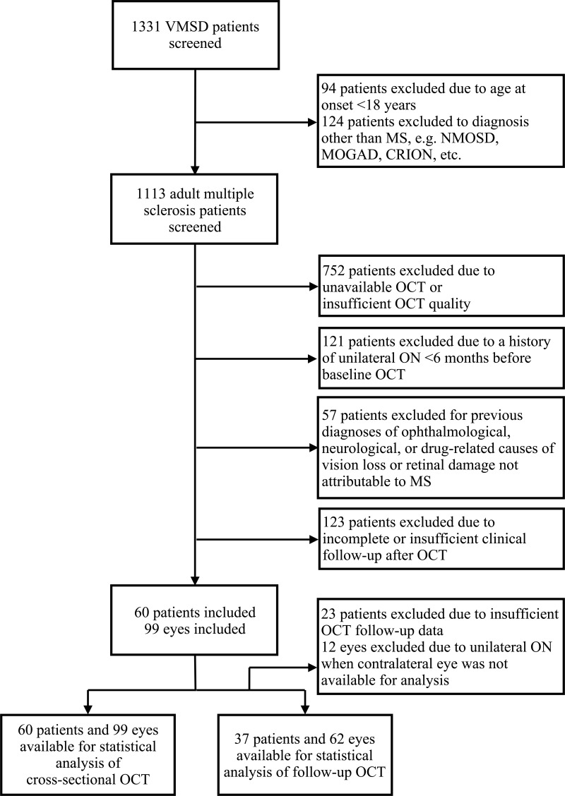
Patients and methods: For this longitudinal observational study, we included MS patients with spectral-domain OCT scans available and ≥1 year of clinical follow-up. The value of peripapillary retinal nerve fiber layer (pRNFL), macular ganglion-cell-and-inner-plexiform-layer (GCIPL) and inner nuclear layer (INL) thickness for prediction of disability worsening and relapse during the observation period was tested by multivariate models.
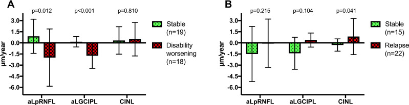
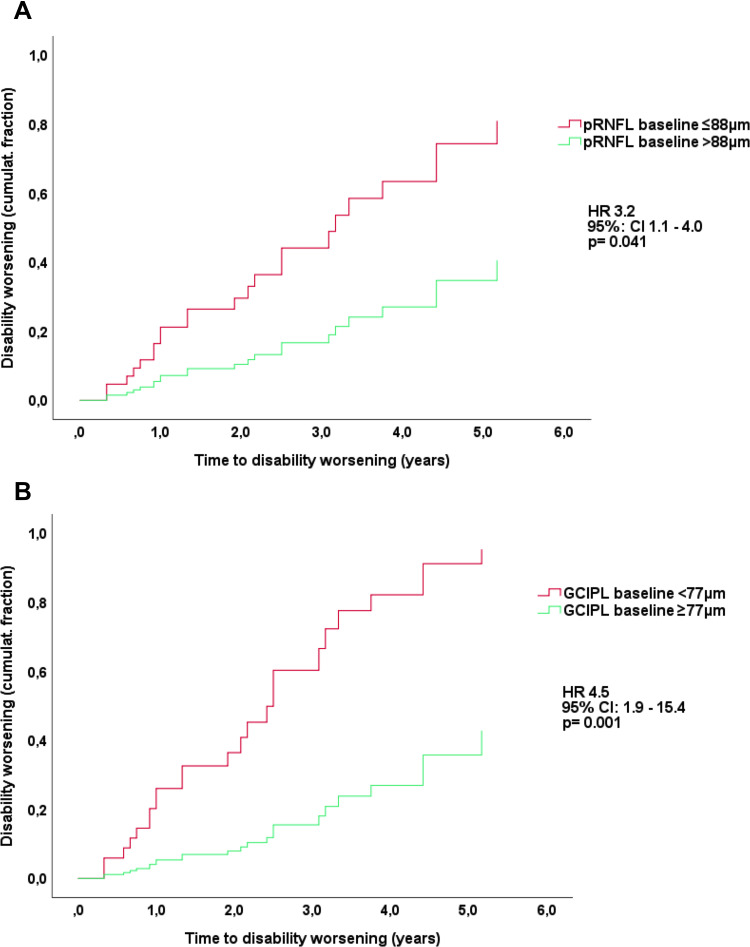
Results: We analyzed 60 MS patients during a mean observation period of 2.9 years (SD 1.8). Lower baseline thickness of GCIPL (cut-off <77µm; HR 4.1, p=0.001) and pRNFL (cut-off ≤88µm; HR 3.1, p=0.019) were associated with an increased risk of disability worsening. Longitudinally, mean thinning rates were -0.8µm/year (SD 1.6) for GCIPL, -0.6µm/year (SD 3.5) for pRNFL. GCIPL thinning ≥1.0µm/year and pRNFL >1.5µm/year is associated with higher likelihood of disability worsening (HR 5.7, p=0.009 and HR 6.8, p=0.003, respectively). INL thickened in patients with relapse by a mean 0.9µm while thinning by 0.3µm in patients without relapse (p=0.04). In multivariate analyses, INL thickening was associated with an increased probability of relapse (OR 17.8, p=0.023).
Conclusion: Cross-sectional and longitudinal measurement of GCIPL and pRNFL thinning is reliable as a biomarker of disability worsening in a real-world setting. Change of INL thickness is a promising marker of relapse, i.e. inflammatory activity.
期刊介绍:
Eye and Brain is an international, peer-reviewed, open access journal focusing on basic research, clinical findings, and expert reviews in the field of visual science and neuro-ophthalmology. The journal’s unique focus is the link between two well-known visual centres, the eye and the brain, with an emphasis on the importance of such connections. All aspects of clinical and especially basic research on the visual system are addressed within the journal as well as significant future directions in vision research and therapeutic measures. This unique journal focuses on neurological aspects of vision – both physiological and pathological. The scope of the journal spans from the cornea to the associational visual cortex and all the visual centers in between. Topics range from basic biological mechanisms to therapeutic treatment, from simple organisms to humans, and utilizing techniques from molecular biology to behavior. The journal especially welcomes primary research articles or review papers that make the connection between the eye and the brain. Specific areas covered in the journal include: Physiology and pathophysiology of visual centers, Eye movement disorders and strabismus, Cellular, biochemical, and molecular features of the visual system, Structural and functional organization of the eye and of the visual cortex, Metabolic demands of the visual system, Diseases and disorders with neuro-ophthalmic manifestations, Clinical and experimental neuro-ophthalmology and visual system pathologies, Epidemiological studies.

 求助内容:
求助内容: 应助结果提醒方式:


