Differences in mandibular condyle and glenoid fossa morphology in relation to vertical and sagittal skeletal patterns: A cone-beam computed tomography study.
{"title":"Differences in mandibular condyle and glenoid fossa morphology in relation to vertical and sagittal skeletal patterns: A cone-beam computed tomography study.","authors":"Kyoung Jin Noh, Hyoung-Seon Baik, Sang-Sun Han, Woowon Jang, Yoon Jeong Choi","doi":"10.4041/kjod.2021.51.2.126","DOIUrl":null,"url":null,"abstract":"<p><strong>Objective: </strong>This study aimed to evaluate the following null hypothesis: there are no differences in the morphology of the temporomandibular joint (TMJ) structures in relation to vertical and sagittal cephalometric patterns.</p><p><strong>Methods: </strong>This retrospective study was performed with 131 participants showing no TMJ symptoms. The participants were divided into Class I, II, and III groups on the basis of their sagittal cephalometric relationships and into hyperdivergent, normodivergent, and hypodivergent groups on the basis of their vertical cephalometric relationships. The following measurements were performed using cone-beam computed tomography images and compared among the groups: condylar volume, condylar size (width, length, and height), fossa size (length and height), and condyle-to-fossa joint spaces at the anterior, superior, and posterior condylar poles.</p><p><strong>Results: </strong>The null hypothesis was rejected. The Class III group showed larger values for condylar width, condylar height, and fossa height than the Class II group (<i>p</i> < 0.05). Condylar volume and superior joint space in the hyperdivergent group were significantly smaller than those in the other two vertical groups (<i>p</i> < 0.001), whereas fossa length and height were significantly larger in the hyperdivergent group than in the other groups (<i>p</i> < 0.01). The hypodivergent group showed a greater condylar width than the hyperdivergent group (<i>p</i> < 0.01). The sagittal and vertical cephalometric patterns showed statistically significant interactions for fossa length and height.</p><p><strong>Conclusions: </strong>TMJ morphology differed across diverse skeletal cephalometric patterns. The fossa length and height were affected by the interactions of the vertical and sagittal skeletal patterns.</p>","PeriodicalId":49934,"journal":{"name":"Korean Journal of Orthodontics","volume":"51 2","pages":"126-134"},"PeriodicalIF":1.9000,"publicationDate":"2021-03-25","publicationTypes":"Journal Article","fieldsOfStudy":null,"isOpenAccess":false,"openAccessPdf":"https://ftp.ncbi.nlm.nih.gov/pub/pmc/oa_pdf/04/86/kjod-51-2-126.PMC7940806.pdf","citationCount":"9","resultStr":null,"platform":"Semanticscholar","paperid":null,"PeriodicalName":"Korean Journal of Orthodontics","FirstCategoryId":"3","ListUrlMain":"https://doi.org/10.4041/kjod.2021.51.2.126","RegionNum":3,"RegionCategory":"医学","ArticlePicture":[],"TitleCN":null,"AbstractTextCN":null,"PMCID":null,"EPubDate":"","PubModel":"","JCR":"Q1","JCRName":"Dentistry","Score":null,"Total":0}
引用次数: 9
Abstract
Objective: This study aimed to evaluate the following null hypothesis: there are no differences in the morphology of the temporomandibular joint (TMJ) structures in relation to vertical and sagittal cephalometric patterns.
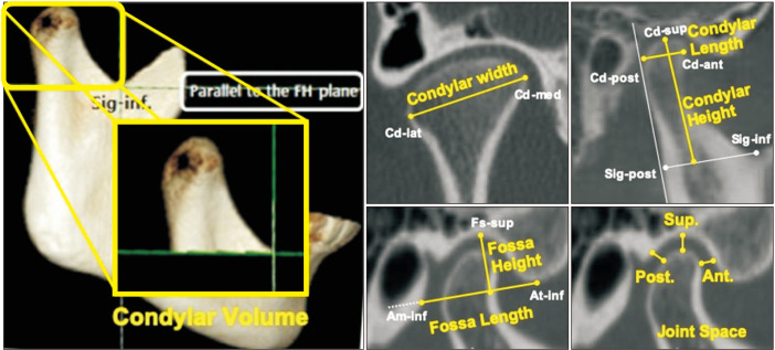
Methods: This retrospective study was performed with 131 participants showing no TMJ symptoms. The participants were divided into Class I, II, and III groups on the basis of their sagittal cephalometric relationships and into hyperdivergent, normodivergent, and hypodivergent groups on the basis of their vertical cephalometric relationships. The following measurements were performed using cone-beam computed tomography images and compared among the groups: condylar volume, condylar size (width, length, and height), fossa size (length and height), and condyle-to-fossa joint spaces at the anterior, superior, and posterior condylar poles.
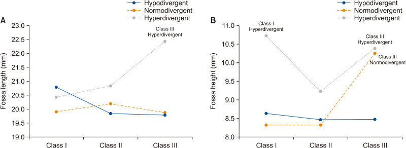
Results: The null hypothesis was rejected. The Class III group showed larger values for condylar width, condylar height, and fossa height than the Class II group (p < 0.05). Condylar volume and superior joint space in the hyperdivergent group were significantly smaller than those in the other two vertical groups (p < 0.001), whereas fossa length and height were significantly larger in the hyperdivergent group than in the other groups (p < 0.01). The hypodivergent group showed a greater condylar width than the hyperdivergent group (p < 0.01). The sagittal and vertical cephalometric patterns showed statistically significant interactions for fossa length and height.
Conclusions: TMJ morphology differed across diverse skeletal cephalometric patterns. The fossa length and height were affected by the interactions of the vertical and sagittal skeletal patterns.
期刊介绍:
The Korean Journal of Orthodontics (KJO) is an international, open access, peer reviewed journal published in January, March, May, July, September, and November each year. It was first launched in 1970 and, as the official scientific publication of Korean Association of Orthodontists, KJO aims to publish high quality clinical and scientific original research papers in all areas related to orthodontics and dentofacial orthopedics. Specifically, its interest focuses on evidence-based investigations of contemporary diagnostic procedures and treatment techniques, expanding to significant clinical reports of diverse treatment approaches.
The scope of KJO covers all areas of orthodontics and dentofacial orthopedics including successful diagnostic procedures and treatment planning, growth and development of the face and its clinical implications, appliance designs, biomechanics, TMJ disorders and adult treatment. Specifically, its latest interest focuses on skeletal anchorage devices, orthodontic appliance and biomaterials, 3 dimensional imaging techniques utilized for dentofacial diagnosis and treatment planning, and orthognathic surgery to correct skeletal disharmony in association of orthodontic treatment.
 求助内容:
求助内容: 应助结果提醒方式:


