Evaluation of a multi-stage convolutional neural network-based fully automated landmark identification system using cone-beam computed tomographysynthesized posteroanterior cephalometric images.
Min-Jung Kim, Yi Liu, Song Hee Oh, Hyo-Won Ahn, Seong-Hun Kim, Gerald Nelson
{"title":"Evaluation of a multi-stage convolutional neural network-based fully automated landmark identification system using cone-beam computed tomographysynthesized posteroanterior cephalometric images.","authors":"Min-Jung Kim, Yi Liu, Song Hee Oh, Hyo-Won Ahn, Seong-Hun Kim, Gerald Nelson","doi":"10.4041/kjod.2021.51.2.77","DOIUrl":null,"url":null,"abstract":"<p><strong>Objective: </strong>To evaluate the accuracy of a multi-stage convolutional neural network (CNN) model-based automated identification system for posteroanterior (PA) cephalometric landmarks.</p><p><strong>Methods: </strong>The multi-stage CNN model was implemented with a personal computer. A total of 430 PA-cephalograms synthesized from cone-beam computed tomography scans (CBCT-PA) were selected as samples. Twenty-three landmarks used for Tweemac analysis were manually identified on all CBCT-PA images by a single examiner. Intra-examiner reproducibility was confirmed by repeating the identification on 85 randomly selected images, which were subsequently set as test data, with a two-week interval before training. For initial learning stage of the multi-stage CNN model, the data from 345 of 430 CBCT-PA images were used, after which the multi-stage CNN model was tested with previous 85 images. The first manual identification on these 85 images was set as a truth ground. The mean radial error (MRE) and successful detection rate (SDR) were calculated to evaluate the errors in manual identification and artificial intelligence (AI) prediction.</p><p><strong>Results: </strong>The AI showed an average MRE of 2.23 ± 2.02 mm with an SDR of 60.88% for errors of 2 mm or lower. However, in a comparison of the repetitive task, the AI predicted landmarks at the same position, while the MRE for the repeated manual identification was 1.31 ± 0.94 mm.</p><p><strong>Conclusions: </strong>Automated identification for CBCT-synthesized PA cephalometric landmarks did not sufficiently achieve the clinically favorable error range of less than 2 mm. However, AI landmark identification on PA cephalograms showed better consistency than manual identification.</p>","PeriodicalId":49934,"journal":{"name":"Korean Journal of Orthodontics","volume":"51 2","pages":"77-85"},"PeriodicalIF":1.9000,"publicationDate":"2021-03-25","publicationTypes":"Journal Article","fieldsOfStudy":null,"isOpenAccess":false,"openAccessPdf":"https://ftp.ncbi.nlm.nih.gov/pub/pmc/oa_pdf/7e/8c/kjod-51-2-77.PMC7940808.pdf","citationCount":"8","resultStr":null,"platform":"Semanticscholar","paperid":null,"PeriodicalName":"Korean Journal of Orthodontics","FirstCategoryId":"3","ListUrlMain":"https://doi.org/10.4041/kjod.2021.51.2.77","RegionNum":3,"RegionCategory":"医学","ArticlePicture":[],"TitleCN":null,"AbstractTextCN":null,"PMCID":null,"EPubDate":"","PubModel":"","JCR":"Q1","JCRName":"Dentistry","Score":null,"Total":0}
引用次数: 8
Abstract
Objective: To evaluate the accuracy of a multi-stage convolutional neural network (CNN) model-based automated identification system for posteroanterior (PA) cephalometric landmarks.
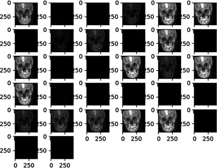
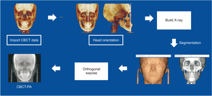
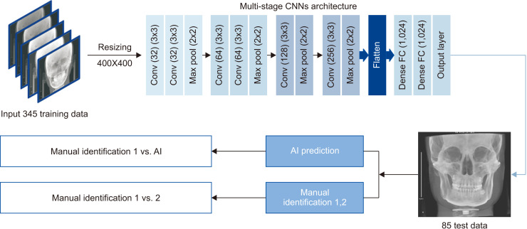
Methods: The multi-stage CNN model was implemented with a personal computer. A total of 430 PA-cephalograms synthesized from cone-beam computed tomography scans (CBCT-PA) were selected as samples. Twenty-three landmarks used for Tweemac analysis were manually identified on all CBCT-PA images by a single examiner. Intra-examiner reproducibility was confirmed by repeating the identification on 85 randomly selected images, which were subsequently set as test data, with a two-week interval before training. For initial learning stage of the multi-stage CNN model, the data from 345 of 430 CBCT-PA images were used, after which the multi-stage CNN model was tested with previous 85 images. The first manual identification on these 85 images was set as a truth ground. The mean radial error (MRE) and successful detection rate (SDR) were calculated to evaluate the errors in manual identification and artificial intelligence (AI) prediction.
Results: The AI showed an average MRE of 2.23 ± 2.02 mm with an SDR of 60.88% for errors of 2 mm or lower. However, in a comparison of the repetitive task, the AI predicted landmarks at the same position, while the MRE for the repeated manual identification was 1.31 ± 0.94 mm.
Conclusions: Automated identification for CBCT-synthesized PA cephalometric landmarks did not sufficiently achieve the clinically favorable error range of less than 2 mm. However, AI landmark identification on PA cephalograms showed better consistency than manual identification.
期刊介绍:
The Korean Journal of Orthodontics (KJO) is an international, open access, peer reviewed journal published in January, March, May, July, September, and November each year. It was first launched in 1970 and, as the official scientific publication of Korean Association of Orthodontists, KJO aims to publish high quality clinical and scientific original research papers in all areas related to orthodontics and dentofacial orthopedics. Specifically, its interest focuses on evidence-based investigations of contemporary diagnostic procedures and treatment techniques, expanding to significant clinical reports of diverse treatment approaches.
The scope of KJO covers all areas of orthodontics and dentofacial orthopedics including successful diagnostic procedures and treatment planning, growth and development of the face and its clinical implications, appliance designs, biomechanics, TMJ disorders and adult treatment. Specifically, its latest interest focuses on skeletal anchorage devices, orthodontic appliance and biomaterials, 3 dimensional imaging techniques utilized for dentofacial diagnosis and treatment planning, and orthognathic surgery to correct skeletal disharmony in association of orthodontic treatment.
 求助内容:
求助内容: 应助结果提醒方式:


