{"title":"Prognostic Value of Electrical Impedance Spectroscopy (EIS) When Used as an Adjunct to Colposcopy - A Longitudinal Study.","authors":"B H Brown, P E Highfield, J A Tidy","doi":"10.2478/joeb-2020-0012","DOIUrl":null,"url":null,"abstract":"<p><strong>Objective: </strong>Colposcopy can be used with Electrical Impedance Spectroscopy (EIS) as an adjunct, to assess the presence of High Grade Cervical Intra-epithelial Neoplasia (CIN2+). This analysis of longitudinal data has used the results from women with a negative colposcopy, in order to see if the initial (index) EIS results were able to predict the women who subsequently developed CIN2+. A further objective was to investigate what tissue structural changes might be reflected in the electrical impedance spectra.</p><p><strong>Methods: </strong>847 patients were referred with low grade cytologly. EIS measurements were made around the transformation zone of the cervix during colposcopy. Every EIS spectrum was matched to a template representing CIN2+ and the result was positive if the match exceeded a probability index threshold. The colposcopic impression was also recorded. All the women who developed biopsy proven CIN2+ within three years of the index colposcopy were identified.</p><p><strong>Results: </strong>The median follow-up was 30.5 months. Where both CI and EIS were initially positive, there was an increased prevalence (8.13%) of CIN2+ developing as opposed to 3.45% in the remaining patients (p=0.0159). In addition, if three or more EIS spectra were positive there was a higher prevalence (9.62% as opposed to 3.56% p=0.0132) of CIN2+ at three years. The index spectra recorded from the women who developed CIN2+ showed EIS changes consistent with increases in the extracellular volume and in cell size inhomogeneity.</p><p><strong>Conclusion: </strong>EIS does offer prognostic information on the risk of CIN2+ developing over the three-year period following the EIS measurements. The changes in EIS spectra are consistent with an increase in cell size diversity as pre-malignancy develops. These changes may be a consequence of increased genetic diversity as neoplasia develops.</p>","PeriodicalId":38125,"journal":{"name":"Journal of Electrical Bioimpedance","volume":" ","pages":"81-86"},"PeriodicalIF":0.0000,"publicationDate":"2020-11-06","publicationTypes":"Journal Article","fieldsOfStudy":null,"isOpenAccess":false,"openAccessPdf":"https://www.ncbi.nlm.nih.gov/pmc/articles/PMC7851983/pdf/","citationCount":"6","resultStr":null,"platform":"Semanticscholar","paperid":null,"PeriodicalName":"Journal of Electrical Bioimpedance","FirstCategoryId":"1085","ListUrlMain":"https://doi.org/10.2478/joeb-2020-0012","RegionNum":0,"RegionCategory":null,"ArticlePicture":[],"TitleCN":null,"AbstractTextCN":null,"PMCID":null,"EPubDate":"2020/1/1 0:00:00","PubModel":"eCollection","JCR":"Q3","JCRName":"Biochemistry, Genetics and Molecular Biology","Score":null,"Total":0}
引用次数: 6
Abstract
Objective: Colposcopy can be used with Electrical Impedance Spectroscopy (EIS) as an adjunct, to assess the presence of High Grade Cervical Intra-epithelial Neoplasia (CIN2+). This analysis of longitudinal data has used the results from women with a negative colposcopy, in order to see if the initial (index) EIS results were able to predict the women who subsequently developed CIN2+. A further objective was to investigate what tissue structural changes might be reflected in the electrical impedance spectra.
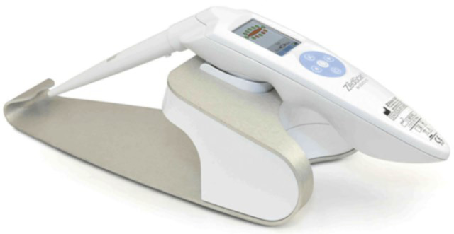
Methods: 847 patients were referred with low grade cytologly. EIS measurements were made around the transformation zone of the cervix during colposcopy. Every EIS spectrum was matched to a template representing CIN2+ and the result was positive if the match exceeded a probability index threshold. The colposcopic impression was also recorded. All the women who developed biopsy proven CIN2+ within three years of the index colposcopy were identified.
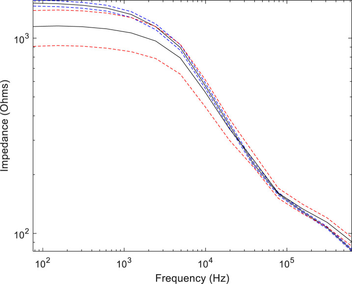
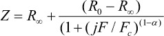
Results: The median follow-up was 30.5 months. Where both CI and EIS were initially positive, there was an increased prevalence (8.13%) of CIN2+ developing as opposed to 3.45% in the remaining patients (p=0.0159). In addition, if three or more EIS spectra were positive there was a higher prevalence (9.62% as opposed to 3.56% p=0.0132) of CIN2+ at three years. The index spectra recorded from the women who developed CIN2+ showed EIS changes consistent with increases in the extracellular volume and in cell size inhomogeneity.
Conclusion: EIS does offer prognostic information on the risk of CIN2+ developing over the three-year period following the EIS measurements. The changes in EIS spectra are consistent with an increase in cell size diversity as pre-malignancy develops. These changes may be a consequence of increased genetic diversity as neoplasia develops.

 求助内容:
求助内容: 应助结果提醒方式:


