A Rare Case of GATA3 Positivity in Pleomorphic Lung Carcinoma in a Patient with History of Intracystic Papillary Carcinoma of the Breast: Primary Lung or Metastatic Disease?
{"title":"A Rare Case of GATA3 Positivity in Pleomorphic Lung Carcinoma in a Patient with History of Intracystic Papillary Carcinoma of the Breast: Primary Lung or Metastatic Disease?","authors":"Evi Abada","doi":"10.1155/2021/6664804","DOIUrl":null,"url":null,"abstract":"<p><p>Pleomorphic lung carcinoma is a rare and aggressive neoplasm accounting for <1% of all lung tumors. It is more common in men and consists of spindle and/or giant cells with an epithelial component. In patients with known histories of malignancies at other sites, diagnosis of a new lung lesion may prove challenging with respect to classification as either primary or metastatic disease, especially in cases with overlapping immunohistochemical staining patterns. This was a case of a 67-year-old female with a newly discovered 1.5 cm nodule in her left lower lung lobe. Her past medical history was significant for an intracystic papillary carcinoma of the right breast diagnosed 8 years prior. Histopathologic examination of the new lung lesion revealed highly pleomorphic cells composed predominantly of neoplastic giant cells and atypical mitotic figures, with geographic areas of necrosis. However, no areas reminiscent of intracystic papillary carcinoma or other forms of breast carcinoma were seen. Immunohistochemistry showed that the tumor cells were immunoreactive for GATA3, TTF1, and napsin A and nonimmunoreactive for p40. Therefore, although this index lung tumor did show positivity with GATA3 staining, it was morphologically different from her original intracystic papillary carcinoma of the breast. In addition, intracystic papillary carcinomas are known to rarely metastasize to other organs, and GATA3 staining has been rarely reported in lung carcinomas. In summary, this case typifies the overlapping immunohistochemical staining patterns that may be seen in different tumors and the role of histopathologic morphology in arriving at the correct diagnosis.</p>","PeriodicalId":45638,"journal":{"name":"Case Reports in Pathology","volume":"2021 ","pages":"6664804"},"PeriodicalIF":0.7000,"publicationDate":"2021-01-20","publicationTypes":"Journal Article","fieldsOfStudy":null,"isOpenAccess":false,"openAccessPdf":"https://www.ncbi.nlm.nih.gov/pmc/articles/PMC7840265/pdf/","citationCount":"0","resultStr":null,"platform":"Semanticscholar","paperid":null,"PeriodicalName":"Case Reports in Pathology","FirstCategoryId":"1085","ListUrlMain":"https://doi.org/10.1155/2021/6664804","RegionNum":0,"RegionCategory":null,"ArticlePicture":[],"TitleCN":null,"AbstractTextCN":null,"PMCID":null,"EPubDate":"2021/1/1 0:00:00","PubModel":"eCollection","JCR":"Q4","JCRName":"PATHOLOGY","Score":null,"Total":0}
引用次数: 0
Abstract
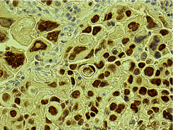
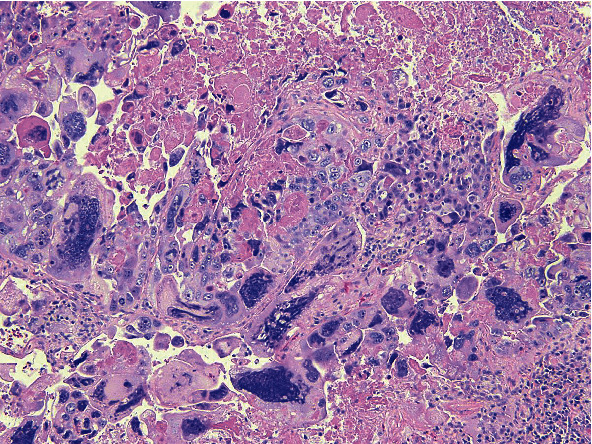
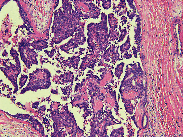
Pleomorphic lung carcinoma is a rare and aggressive neoplasm accounting for <1% of all lung tumors. It is more common in men and consists of spindle and/or giant cells with an epithelial component. In patients with known histories of malignancies at other sites, diagnosis of a new lung lesion may prove challenging with respect to classification as either primary or metastatic disease, especially in cases with overlapping immunohistochemical staining patterns. This was a case of a 67-year-old female with a newly discovered 1.5 cm nodule in her left lower lung lobe. Her past medical history was significant for an intracystic papillary carcinoma of the right breast diagnosed 8 years prior. Histopathologic examination of the new lung lesion revealed highly pleomorphic cells composed predominantly of neoplastic giant cells and atypical mitotic figures, with geographic areas of necrosis. However, no areas reminiscent of intracystic papillary carcinoma or other forms of breast carcinoma were seen. Immunohistochemistry showed that the tumor cells were immunoreactive for GATA3, TTF1, and napsin A and nonimmunoreactive for p40. Therefore, although this index lung tumor did show positivity with GATA3 staining, it was morphologically different from her original intracystic papillary carcinoma of the breast. In addition, intracystic papillary carcinomas are known to rarely metastasize to other organs, and GATA3 staining has been rarely reported in lung carcinomas. In summary, this case typifies the overlapping immunohistochemical staining patterns that may be seen in different tumors and the role of histopathologic morphology in arriving at the correct diagnosis.

 求助内容:
求助内容: 应助结果提醒方式:


