Gabriela Edwards-Faret, Karina González-Pinto, Arantxa Cebrián-Silla, Johany Peñailillo, José Manuel García-Verdugo, Juan Larraín
{"title":"Cellular response to spinal cord injury in regenerative and non-regenerative stages in Xenopus laevis.","authors":"Gabriela Edwards-Faret, Karina González-Pinto, Arantxa Cebrián-Silla, Johany Peñailillo, José Manuel García-Verdugo, Juan Larraín","doi":"10.1186/s13064-021-00152-2","DOIUrl":null,"url":null,"abstract":"<p><strong>Background: </strong>The efficient regenerative abilities at larvae stages followed by a non-regenerative response after metamorphosis in froglets makes Xenopus an ideal model organism to understand the cellular responses leading to spinal cord regeneration.</p><p><strong>Methods: </strong>We compared the cellular response to spinal cord injury between the regenerative and non-regenerative stages of Xenopus laevis. For this analysis, we used electron microscopy, immunofluorescence and histological staining of the extracellular matrix. We generated two transgenic lines: i) the reporter line with the zebrafish GFAP regulatory regions driving the expression of EGFP, and ii) a cell specific inducible ablation line with the same GFAP regulatory regions. In addition, we used FACS to isolate EGFP<sup>+</sup> cells for RNAseq analysis.</p><p><strong>Results: </strong>In regenerative stage animals, spinal cord regeneration triggers a rapid sealing of the injured stumps, followed by proliferation of cells lining the central canal, and formation of rosette-like structures in the ablation gap. In addition, the central canal is filled by cells with similar morphology to the cells lining the central canal, neurons, axons, and even synaptic structures. Regeneration is almost completed after 20 days post injury. In non-regenerative stage animals, mostly damaged tissue was observed, without clear closure of the stumps. The ablation gap was filled with fibroblast-like cells, and deposition of extracellular matrix components. No reconstruction of the spinal cord was observed even after 40 days post injury. Cellular markers analysis confirmed these histological differences, a transient increase of vimentin, fibronectin and collagen was detected in regenerative stages, contrary to a sustained accumulation of most of these markers, including chondroitin sulfate proteoglycans in the NR-stage. The zebrafish GFAP transgenic line was validated, and we have demonstrated that is a very reliable and new tool to study the role of neural stem progenitor cells (NSPCs). RNASeq of GFAP::EGFP cells has allowed us to clearly demonstrate that indeed these cells are NSPCs. On the contrary, the GFAP::EGFP transgene is mainly expressed in astrocytes in non-regenerative stages. During regenerative stages, spinal cord injury activates proliferation of NSPCs, and we found that are mainly differentiated into neurons and glial cells. Specific ablation of these cells abolished proper regeneration, confirming that NSPCs cells are necessary for functional regeneration of the spinal cord.</p><p><strong>Conclusions: </strong>The cellular response to spinal cord injury in regenerative and non-regenerative stages is profoundly different between both stages. A key hallmark of the regenerative response is the activation of NSPCs, which massively proliferate, and are differentiated into neurons to reconstruct the spinal cord. Also very notably, no glial scar formation is observed in regenerative stages, but a transient, glial scar-like structure is formed in non-regenerative stage animals.</p>","PeriodicalId":49764,"journal":{"name":"Neural Development","volume":" ","pages":"2"},"PeriodicalIF":2.5000,"publicationDate":"2021-02-02","publicationTypes":"Journal Article","fieldsOfStudy":null,"isOpenAccess":false,"openAccessPdf":"https://www.ncbi.nlm.nih.gov/pmc/articles/PMC7852093/pdf/","citationCount":"14","resultStr":null,"platform":"Semanticscholar","paperid":null,"PeriodicalName":"Neural Development","FirstCategoryId":"99","ListUrlMain":"https://doi.org/10.1186/s13064-021-00152-2","RegionNum":3,"RegionCategory":"生物学","ArticlePicture":[],"TitleCN":null,"AbstractTextCN":null,"PMCID":null,"EPubDate":"","PubModel":"","JCR":"Q1","JCRName":"DEVELOPMENTAL BIOLOGY","Score":null,"Total":0}
引用次数: 14
Abstract
Background: The efficient regenerative abilities at larvae stages followed by a non-regenerative response after metamorphosis in froglets makes Xenopus an ideal model organism to understand the cellular responses leading to spinal cord regeneration.
Methods: We compared the cellular response to spinal cord injury between the regenerative and non-regenerative stages of Xenopus laevis. For this analysis, we used electron microscopy, immunofluorescence and histological staining of the extracellular matrix. We generated two transgenic lines: i) the reporter line with the zebrafish GFAP regulatory regions driving the expression of EGFP, and ii) a cell specific inducible ablation line with the same GFAP regulatory regions. In addition, we used FACS to isolate EGFP+ cells for RNAseq analysis.
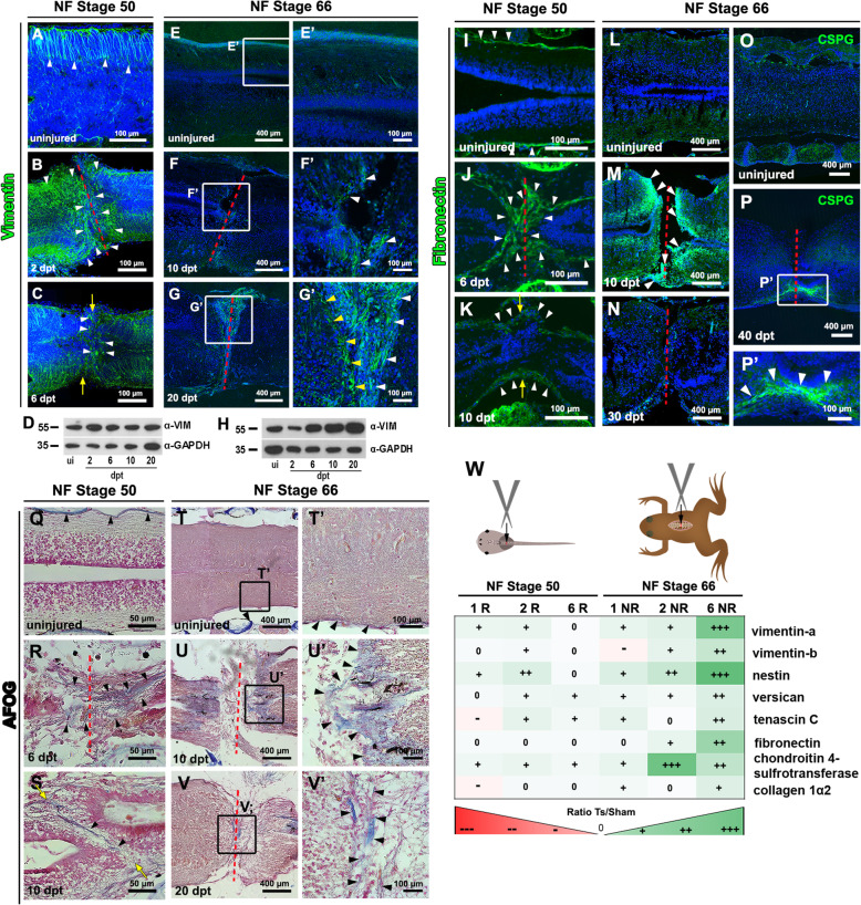
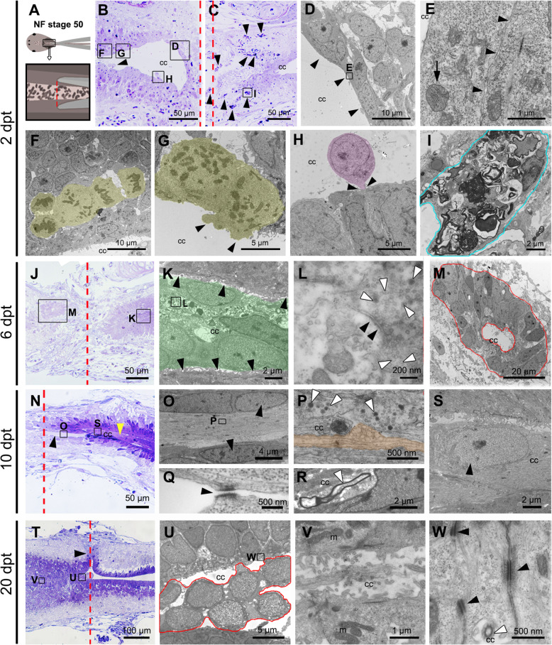
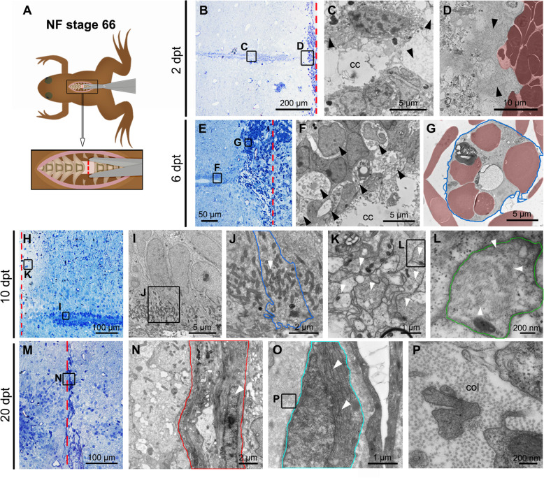
Results: In regenerative stage animals, spinal cord regeneration triggers a rapid sealing of the injured stumps, followed by proliferation of cells lining the central canal, and formation of rosette-like structures in the ablation gap. In addition, the central canal is filled by cells with similar morphology to the cells lining the central canal, neurons, axons, and even synaptic structures. Regeneration is almost completed after 20 days post injury. In non-regenerative stage animals, mostly damaged tissue was observed, without clear closure of the stumps. The ablation gap was filled with fibroblast-like cells, and deposition of extracellular matrix components. No reconstruction of the spinal cord was observed even after 40 days post injury. Cellular markers analysis confirmed these histological differences, a transient increase of vimentin, fibronectin and collagen was detected in regenerative stages, contrary to a sustained accumulation of most of these markers, including chondroitin sulfate proteoglycans in the NR-stage. The zebrafish GFAP transgenic line was validated, and we have demonstrated that is a very reliable and new tool to study the role of neural stem progenitor cells (NSPCs). RNASeq of GFAP::EGFP cells has allowed us to clearly demonstrate that indeed these cells are NSPCs. On the contrary, the GFAP::EGFP transgene is mainly expressed in astrocytes in non-regenerative stages. During regenerative stages, spinal cord injury activates proliferation of NSPCs, and we found that are mainly differentiated into neurons and glial cells. Specific ablation of these cells abolished proper regeneration, confirming that NSPCs cells are necessary for functional regeneration of the spinal cord.
Conclusions: The cellular response to spinal cord injury in regenerative and non-regenerative stages is profoundly different between both stages. A key hallmark of the regenerative response is the activation of NSPCs, which massively proliferate, and are differentiated into neurons to reconstruct the spinal cord. Also very notably, no glial scar formation is observed in regenerative stages, but a transient, glial scar-like structure is formed in non-regenerative stage animals.
期刊介绍:
Neural Development is a peer-reviewed open access, online journal, which features studies that use molecular, cellular, physiological or behavioral methods to provide novel insights into the mechanisms that underlie the formation of the nervous system.
Neural Development aims to discover how the nervous system arises and acquires the abilities to sense the world and control adaptive motor output. The field includes analysis of how progenitor cells form a nervous system during embryogenesis, and how the initially formed neural circuits are shaped by experience during early postnatal life. Some studies use well-established, genetically accessible model systems, but valuable insights are also obtained from less traditional models that provide behavioral or evolutionary insights.

 求助内容:
求助内容: 应助结果提醒方式:


