Ben A. Weissman, Enmei Niu, Renshan Ge, Chantal M. Sottas, Michael Holmes, James C. Hutson, Matthew P. Hardy
{"title":"Paracrine Modulation of Androgen Synthesis in Rat Leydig Cells by Nitric Oxide","authors":"Ben A. Weissman, Enmei Niu, Renshan Ge, Chantal M. Sottas, Michael Holmes, James C. Hutson, Matthew P. Hardy","doi":"10.2164/jandrol.04178","DOIUrl":null,"url":null,"abstract":"<p><b>ABSTRACT: </b> The free radical nitric oxide (NO), generated through the oxidation of <span>l</span>-arginine to <span>l</span>-citrulline by NO synthases (NOSs), has been shown to inhibit steroidogenic pathways. NOS isoforms are known to be present in rat and human testes. Our study examined the sensitivity of Leydig cells to NO and determined whether NOS activity resides in Leydig cells or in another cell type such as the testicular macrophage. The results showed a low level of <span>l</span>-[<sup>14</sup>C]arginine conversion in purified rat Leydig cell homogenates. Administration of the NOS inhibitor L-N<sup>G</sup>-nitro-arginine methyl ester (L-NAME), or the calcium chelator ethylenebis (oxyethylenenitrilo)tetraacetic acid (EGTA), had no effect on <span>l</span>-[<sup>14</sup>C]citrulline accumulation. Increased intracellular Ca<sup>2+</sup> concentrations that were induced by a calcium ionophore, or the addition of luteinizing hormone (LH), failed to affect NO formation in intact cells that were cultured in vitro. Introduction of a high concentration of the NO precursor <span>l</span>-arginine did not decrease testosterone (T) production, and NOS inhibitors did not increase T biosynthesis. However, exposing Leydig cells to low concentrations of the NO donor S-nitrosoglutathione (GSNO) induced a dramatic blockade of T production under basal and LH-stimulated conditions. DNA array assays showed a low level of expression of endothelial NOS (eNOS), while the neuronal and inducible isoforms of NOS (nNOS and iNOS) were below detection levels. Reverse transcriptase-polymerase chain reaction (RT-PCR) analyses confirmed these findings and demonstrated the presence of high iNOS messenger RNA (mRNA) levels in activated testicular macrophages that produced large amounts of NO. These data suggest that, while T production in rat Leydig cells is highly sensitive to NO and an endogenous NO-generating system is not present in these cells, NOS activity is more likely to reside in activated testicular macrophages.</p>","PeriodicalId":15029,"journal":{"name":"Journal of andrology","volume":"26 3","pages":"369-378"},"PeriodicalIF":0.0000,"publicationDate":"2013-01-02","publicationTypes":"Journal Article","fieldsOfStudy":null,"isOpenAccess":false,"openAccessPdf":"https://sci-hub-pdf.com/10.2164/jandrol.04178","citationCount":"68","resultStr":null,"platform":"Semanticscholar","paperid":null,"PeriodicalName":"Journal of andrology","FirstCategoryId":"1085","ListUrlMain":"https://onlinelibrary.wiley.com/doi/10.2164/jandrol.04178","RegionNum":0,"RegionCategory":null,"ArticlePicture":[],"TitleCN":null,"AbstractTextCN":null,"PMCID":null,"EPubDate":"","PubModel":"","JCR":"","JCRName":"","Score":null,"Total":0}
引用次数: 68
Abstract
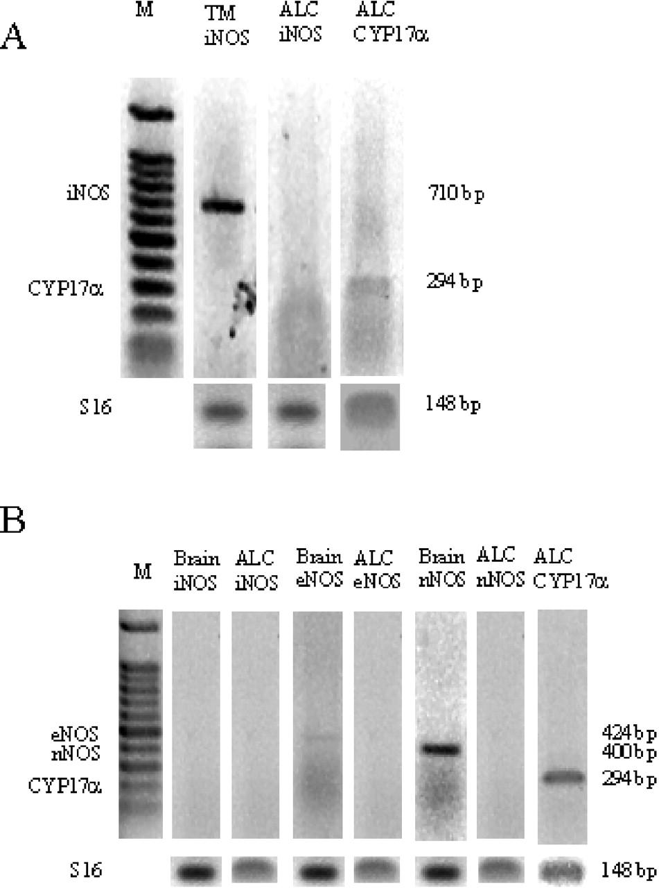
ABSTRACT: The free radical nitric oxide (NO), generated through the oxidation of l-arginine to l-citrulline by NO synthases (NOSs), has been shown to inhibit steroidogenic pathways. NOS isoforms are known to be present in rat and human testes. Our study examined the sensitivity of Leydig cells to NO and determined whether NOS activity resides in Leydig cells or in another cell type such as the testicular macrophage. The results showed a low level of l-[14C]arginine conversion in purified rat Leydig cell homogenates. Administration of the NOS inhibitor L-NG-nitro-arginine methyl ester (L-NAME), or the calcium chelator ethylenebis (oxyethylenenitrilo)tetraacetic acid (EGTA), had no effect on l-[14C]citrulline accumulation. Increased intracellular Ca2+ concentrations that were induced by a calcium ionophore, or the addition of luteinizing hormone (LH), failed to affect NO formation in intact cells that were cultured in vitro. Introduction of a high concentration of the NO precursor l-arginine did not decrease testosterone (T) production, and NOS inhibitors did not increase T biosynthesis. However, exposing Leydig cells to low concentrations of the NO donor S-nitrosoglutathione (GSNO) induced a dramatic blockade of T production under basal and LH-stimulated conditions. DNA array assays showed a low level of expression of endothelial NOS (eNOS), while the neuronal and inducible isoforms of NOS (nNOS and iNOS) were below detection levels. Reverse transcriptase-polymerase chain reaction (RT-PCR) analyses confirmed these findings and demonstrated the presence of high iNOS messenger RNA (mRNA) levels in activated testicular macrophages that produced large amounts of NO. These data suggest that, while T production in rat Leydig cells is highly sensitive to NO and an endogenous NO-generating system is not present in these cells, NOS activity is more likely to reside in activated testicular macrophages.

 求助内容:
求助内容: 应助结果提醒方式:


