Ingrid M. C. de Pauw, Alan K. Goff, Ann van Soom, Steven Verberckmoes, Aart de Kruif
{"title":"Hormonal Regulation of Bovine Secretory Proteins Derived From Caput and Cauda Epididymal Epithelial Cell Cultures","authors":"Ingrid M. C. de Pauw, Alan K. Goff, Ann van Soom, Steven Verberckmoes, Aart de Kruif","doi":"10.1002/j.1939-4640.2003.tb02689.x","DOIUrl":null,"url":null,"abstract":"<p><b>ABSTRACT: </b> The goal of this study was to investigate the effect of hormones (testosterone, dihydrotestosterone [DHT], and hydrocortisone) on the protein secretion of caput and cauda epididymal epithelial cells cultured in principal cell medium (PCM). A confluent monolayer of caput and cauda epididymal epithelial cells was obtained from serum-containing PCM in the presence or absence of hormones after 7 days of culture at 38.5°C (5% CO<sub>2</sub> in air). The protein secretion of epididymal epithelial monolayers incubated in serum-free PCM for 3 days was examined. The secreted proteins were separated by 2-dimensional sodium dodecyl sulfate-polyacrylamide gel electrophoresis (2D SDS-PAGE). A comparison of the different protein patterns showed 61 spots, of which 11 were secreted only in the presence of hormones, 3 appeared to show hormone-related changes, and 25 were region-specific. Most of these secreted proteins were low-molecular-weight acidic proteins. To obtain evidence of the epididymal origin of the secreted proteins, proteins present in caput and cauda epididymal plasma were analyzed. In conclusion, our data indicate that hormones influence the synthesis of a number of caput and cauda epididymal proteins. Some of these proteins could be important for improving our understanding of spermatozoa maturation and storage and their acquisition of fertilizing ability.</p>","PeriodicalId":15029,"journal":{"name":"Journal of andrology","volume":"24 3","pages":"401-407"},"PeriodicalIF":0.0000,"publicationDate":"2013-01-02","publicationTypes":"Journal Article","fieldsOfStudy":null,"isOpenAccess":false,"openAccessPdf":"https://sci-hub-pdf.com/10.1002/j.1939-4640.2003.tb02689.x","citationCount":"14","resultStr":null,"platform":"Semanticscholar","paperid":null,"PeriodicalName":"Journal of andrology","FirstCategoryId":"1085","ListUrlMain":"https://onlinelibrary.wiley.com/doi/10.1002/j.1939-4640.2003.tb02689.x","RegionNum":0,"RegionCategory":null,"ArticlePicture":[],"TitleCN":null,"AbstractTextCN":null,"PMCID":null,"EPubDate":"","PubModel":"","JCR":"","JCRName":"","Score":null,"Total":0}
引用次数: 14
Abstract
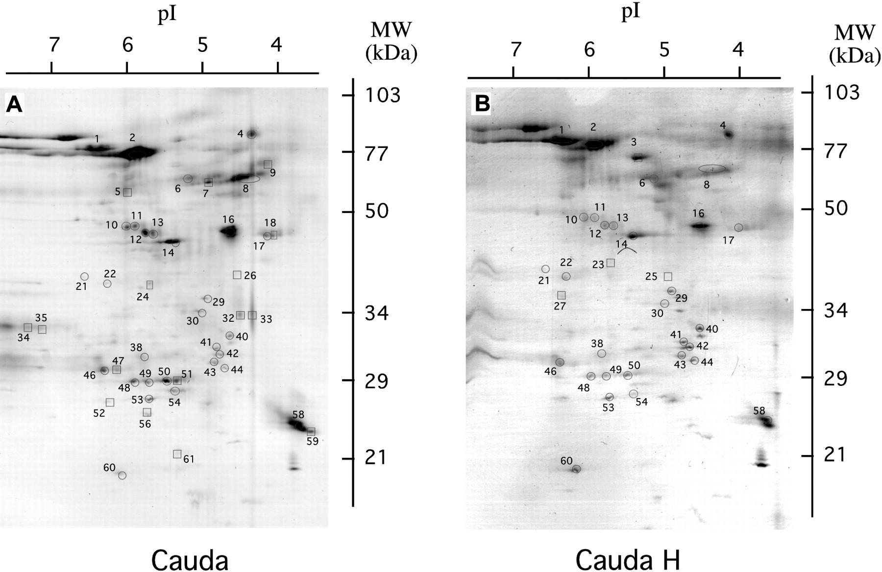
ABSTRACT: The goal of this study was to investigate the effect of hormones (testosterone, dihydrotestosterone [DHT], and hydrocortisone) on the protein secretion of caput and cauda epididymal epithelial cells cultured in principal cell medium (PCM). A confluent monolayer of caput and cauda epididymal epithelial cells was obtained from serum-containing PCM in the presence or absence of hormones after 7 days of culture at 38.5°C (5% CO2 in air). The protein secretion of epididymal epithelial monolayers incubated in serum-free PCM for 3 days was examined. The secreted proteins were separated by 2-dimensional sodium dodecyl sulfate-polyacrylamide gel electrophoresis (2D SDS-PAGE). A comparison of the different protein patterns showed 61 spots, of which 11 were secreted only in the presence of hormones, 3 appeared to show hormone-related changes, and 25 were region-specific. Most of these secreted proteins were low-molecular-weight acidic proteins. To obtain evidence of the epididymal origin of the secreted proteins, proteins present in caput and cauda epididymal plasma were analyzed. In conclusion, our data indicate that hormones influence the synthesis of a number of caput and cauda epididymal proteins. Some of these proteins could be important for improving our understanding of spermatozoa maturation and storage and their acquisition of fertilizing ability.

 求助内容:
求助内容: 应助结果提醒方式:


