Ultrasound molecular imaging of p32 protein translocation for evaluation of tumor metastasis

IF 12.8 1区 医学 Q1 ENGINEERING, BIOMEDICAL
Yongsheng Hao , Jingna Luo , Yuanyuan Wang , Zhenzhou Li , Xiangwei Wang , Fei Yan
{"title":"Ultrasound molecular imaging of p32 protein translocation for evaluation of tumor metastasis","authors":"Yongsheng Hao ,&nbsp;Jingna Luo ,&nbsp;Yuanyuan Wang ,&nbsp;Zhenzhou Li ,&nbsp;Xiangwei Wang ,&nbsp;Fei Yan","doi":"10.1016/j.biomaterials.2022.121974","DOIUrl":null,"url":null,"abstract":"<div><p><span>Protein translocation is an essential process for living cells to respond to different physiological, pathological or environmental stimuli. However, its abnormal occurrence usually results in undesirable outcomes such as tumors. To date, there is still a lack of appropriate methods to detect this event in live animals in a real-time manner. Here, we identified the gradually increased cell-surface translocation of p32 protein from mitochondria during tumor progression. LyP-1-modified gas vesicles (LyP-1-GVs) were developed through conjugating LyP-1 (p32-targeting peptide) to the biosynthetic GVs to monitor the cell-surface level of p32 translocation. The resulting LyP-1-GVs have about 200 nm particle size and good tumor cell targeting performance. Upon systemic administration, LyP-1-GVs can traverse through blood vessels and bind to the tumor cells, producing strong contrast imaging signals in comparison with the non-targeted GVs. The contrast imaging signals correlate well with the cell-surface translocation level of p32 protein and tumor metastatic ability. To our knowledge, this is the first report about the </span><em>in vivo</em><span><span> detection of protein translocation to cell membrane from mitochondria by ultrasound </span>molecular imaging. Our study provides a new strategy to explore the molecular events of protein membrane translocations for evaluation of tumor metastasis at the live animal level.</span></p></div>","PeriodicalId":254,"journal":{"name":"Biomaterials","volume":"293 ","pages":"Article 121974"},"PeriodicalIF":12.8000,"publicationDate":"2023-02-01","publicationTypes":"Journal Article","fieldsOfStudy":null,"isOpenAccess":false,"openAccessPdf":"","citationCount":"0","resultStr":null,"platform":"Semanticscholar","paperid":null,"PeriodicalName":"Biomaterials","FirstCategoryId":"5","ListUrlMain":"https://www.sciencedirect.com/science/article/pii/S0142961222006147","RegionNum":1,"RegionCategory":"医学","ArticlePicture":[],"TitleCN":null,"AbstractTextCN":null,"PMCID":null,"EPubDate":"","PubModel":"","JCR":"Q1","JCRName":"ENGINEERING, BIOMEDICAL","Score":null,"Total":0}
引用次数: 0

Abstract

Protein translocation is an essential process for living cells to respond to different physiological, pathological or environmental stimuli. However, its abnormal occurrence usually results in undesirable outcomes such as tumors. To date, there is still a lack of appropriate methods to detect this event in live animals in a real-time manner. Here, we identified the gradually increased cell-surface translocation of p32 protein from mitochondria during tumor progression. LyP-1-modified gas vesicles (LyP-1-GVs) were developed through conjugating LyP-1 (p32-targeting peptide) to the biosynthetic GVs to monitor the cell-surface level of p32 translocation. The resulting LyP-1-GVs have about 200 nm particle size and good tumor cell targeting performance. Upon systemic administration, LyP-1-GVs can traverse through blood vessels and bind to the tumor cells, producing strong contrast imaging signals in comparison with the non-targeted GVs. The contrast imaging signals correlate well with the cell-surface translocation level of p32 protein and tumor metastatic ability. To our knowledge, this is the first report about the in vivo detection of protein translocation to cell membrane from mitochondria by ultrasound molecular imaging. Our study provides a new strategy to explore the molecular events of protein membrane translocations for evaluation of tumor metastasis at the live animal level.
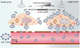
Abstract Image

p32蛋白易位的超声分子成像评价肿瘤转移
蛋白质易位是活细胞对不同生理、病理或环境刺激作出反应的重要过程。然而,它的异常发生通常会导致不良后果,如肿瘤。迄今为止,仍然缺乏在活体动物中实时检测这一事件的适当方法。在这里,我们发现在肿瘤进展过程中,线粒体p32蛋白的细胞表面易位逐渐增加。通过将lpp -1 (p32靶向肽)偶联到生物合成的GVs上,制备了lpp -1修饰的气体囊泡(lypp -1-GVs),以监测p32易位的细胞表面水平。所得LyP-1-GVs粒径约200 nm,具有良好的肿瘤细胞靶向性能。经全身给药后,lyp -1- gv可以穿过血管并与肿瘤细胞结合,与非靶向gv相比,产生强烈的对比成像信号。对比成像信号与p32蛋白的细胞表面易位水平和肿瘤转移能力密切相关。据我们所知,这是第一次用超声分子成像技术在体内检测线粒体向细胞膜转运的蛋白质。我们的研究提供了一种新的策略来探索蛋白质膜易位的分子事件,以评估活体动物水平的肿瘤转移。
本文章由计算机程序翻译,如有差异,请以英文原文为准。
求助全文
约1分钟内获得全文 求助全文
来源期刊
Biomaterials
Biomaterials 工程技术-材料科学:生物材料
CiteScore
26.00
自引率
2.90%
发文量
565
审稿时长
46 days
期刊介绍: Biomaterials is an international journal covering the science and clinical application of biomaterials. A biomaterial is now defined as a substance that has been engineered to take a form which, alone or as part of a complex system, is used to direct, by control of interactions with components of living systems, the course of any therapeutic or diagnostic procedure. It is the aim of the journal to provide a peer-reviewed forum for the publication of original papers and authoritative review and opinion papers dealing with the most important issues facing the use of biomaterials in clinical practice. The scope of the journal covers the wide range of physical, biological and chemical sciences that underpin the design of biomaterials and the clinical disciplines in which they are used. These sciences include polymer synthesis and characterization, drug and gene vector design, the biology of the host response, immunology and toxicology and self assembly at the nanoscale. Clinical applications include the therapies of medical technology and regenerative medicine in all clinical disciplines, and diagnostic systems that reply on innovative contrast and sensing agents. The journal is relevant to areas such as cancer diagnosis and therapy, implantable devices, drug delivery systems, gene vectors, bionanotechnology and tissue engineering.
×
引用
GB/T 7714-2015
复制
MLA
复制
APA
复制
导出至
BibTeX EndNote RefMan NoteFirst NoteExpress
×
提示
您的信息不完整,为了账户安全,请先补充。
现在去补充
×
提示
您因"违规操作"
具体请查看互助需知
我知道了
×
提示
确定
请完成安全验证×
copy
已复制链接
快去分享给好友吧!
我知道了
右上角分享
点击右上角分享
0
联系我们:info@booksci.cn Book学术提供免费学术资源搜索服务,方便国内外学者检索中英文文献。致力于提供最便捷和优质的服务体验。 Copyright © 2023 布克学术 All rights reserved.
京ICP备2023020795号-1
ghs 京公网安备 11010802042870号
Book学术文献互助
Book学术文献互助群
群 号:481959085
Book学术官方微信