[Adipose tissue-derived extracellular vesicles ameliorate zoledronic acid-induced functional impairment of bone marrow mesenchymal stem cells: an in vitro study].
X Y Sun, Z J Pan, Z W Cao, X X Zhou, L Wang, P Y Hong, J Pan
{"title":"[Adipose tissue-derived extracellular vesicles ameliorate zoledronic acid-induced functional impairment of bone marrow mesenchymal stem cells: an <i>in vitro</i> study].","authors":"X Y Sun, Z J Pan, Z W Cao, X X Zhou, L Wang, P Y Hong, J Pan","doi":"10.3760/cma.j.cn112144-20260506-00279","DOIUrl":null,"url":null,"abstract":"<p><p><b>Objective:</b> To investigate the effects of adipose tissue-derived extracellular vesicles (AT-EVs) on zoledronic acid (ZA)-induced impairment of proliferation, migration, osteogenic differentiation and apoptosis of bone marrow mesenchymal stem cells (BMSCs), and to preliminarily explore the underlying molecular mechanisms. <b>Methods:</b> AT-EVs were isolated from subcutaneous adipose tissue of 4-week-old male SPF SD rats, and characterized by transmission electron microscopy, nanoparticle tracking analysis and Western blotting; the uptake of AT-EVs by BMSCs was observed. BMSCs derived from the femurs and tibias of rats of the same batch were isolated and cultured, and identified by flow cytometry and tri-lineage differentiation. The working concentration of ZA was screened by cell counting kit-8 (CCK-8). BMSCs were divided into control, ZA (5 μmol/L), and ZA+AT-EVs (5 μmol/L ZA+50 μg/ml AT-EVs) groups; proliferation was evaluated by CCK-8 combined with 5-ethynyl-2'-deoxyuridine (EdU) incorporation assay, and migration was evaluated by wound healing and Transwell assays; for the apoptosis assay, 10 μmol/L ZA was applied and detected by flow cytometry after Annexin V/PI staining. The osteogenic differentiation experiment included five groups (Control, Osteogenic induction, Osteogenic induction+AT-EVs, ZA, and ZA+AT-EVs), in which alkaline phosphatase (ALP) staining and Alizarin red S staining were performed, mineralized nodule formation was quantified by the cetylpyridinium chloride decolorization method, and the mRNA expression of Runx2, ALP and OCN was detected by real-time quantitative PCR. Cells from the ZA and ZA+AT-EVs groups after 7 days of osteogenic induction were subjected to liquid chromatography-tandem mass spectrometry-based proteomic analysis, followed by principal component analysis, Gene Ontology and Kyoto Encyclopedia of Genes and Genomes enrichment analyses. <b>Results:</b> AT-EVs exhibited a cup-shaped bilayer membrane structure with a peak diameter of 116.8 nm, were positive for CD9, CD63 and CD81 and negative for Calnexin, and were efficiently taken up by BMSCs. CCK-8 results showed that 5 μmol/L ZA significantly inhibited BMSCs proliferation (<i>P</i><0.001), and AT-EVs partially restored the proliferation capacity. The EdU incorporation assay further showed that, compared with the Control group, the EdU-positive rate of the ZA group was significantly decreased (<i>P</i><0.001), while that of the ZA+AT-EVs group was significantly increased compared with the ZA group (<i>P</i><0.01), consistent with the CCK-8 results. Compared with the ZA group, the ZA+AT-EVs group showed enhanced BMSCs migration (all <i>P</i><0.05) and a significantly reduced total apoptosis rate (<i>P</i><0.001). Western blotting further showed that AT-EVs down-regulated the ZA-induced elevation of cleaved caspase-3 (<i>P</i><0.001) and BAX (<i>P</i><0.01) and up-regulated Bcl-2 expression. AT-EVs significantly ameliorated the ZA-induced suppression of BMSCs ALP expression and mineralized nodule formation (<i>P</i><0.001). qPCR showed that AT-EVs up-regulated the ZA-suppressed expression of Runx2, ALP and OCN (<i>P</i><0.05), whereas no significant difference was found between the Osteogenic induction+AT-EVs group and the Osteogenic induction group. Proteomic analysis identified 483 differentially expressed proteins (344 up-regulated and 139 down-regulated), which were mainly enriched in steroid biosynthesis, the cGMP-PKG signaling pathway and cytoskeleton regulation pathways. <b>Conclusions:</b> AT-EVs can effectively ameliorate the ZA-induced impairment of proliferation, migration and osteogenic differentiation of BMSCs and reduce apoptosis in vitro, and their effects may be related to the overall down-regulation of steroid biosynthesis pathway-related proteins and the predominant up-regulation of cGMP-PKG signaling pathway-related proteins in the ZA+AT-EVs group.</p>","PeriodicalId":23965,"journal":{"name":"中华口腔医学杂志","volume":"61 10","pages":"1507-1518"},"PeriodicalIF":0.0000,"publicationDate":"2026-09-30","publicationTypes":"Journal Article","fieldsOfStudy":null,"isOpenAccess":false,"openAccessPdf":"","citationCount":"0","resultStr":null,"platform":"Semanticscholar","paperid":null,"PeriodicalName":"中华口腔医学杂志","FirstCategoryId":"3","ListUrlMain":"https://doi.org/10.3760/cma.j.cn112144-20260506-00279","RegionNum":0,"RegionCategory":null,"ArticlePicture":[],"TitleCN":null,"AbstractTextCN":null,"PMCID":null,"EPubDate":"","PubModel":"","JCR":"Q4","JCRName":"Medicine","Score":null,"Total":0}
引用次数: 0
Abstract
Objective: To investigate the effects of adipose tissue-derived extracellular vesicles (AT-EVs) on zoledronic acid (ZA)-induced impairment of proliferation, migration, osteogenic differentiation and apoptosis of bone marrow mesenchymal stem cells (BMSCs), and to preliminarily explore the underlying molecular mechanisms. Methods: AT-EVs were isolated from subcutaneous adipose tissue of 4-week-old male SPF SD rats, and characterized by transmission electron microscopy, nanoparticle tracking analysis and Western blotting; the uptake of AT-EVs by BMSCs was observed. BMSCs derived from the femurs and tibias of rats of the same batch were isolated and cultured, and identified by flow cytometry and tri-lineage differentiation. The working concentration of ZA was screened by cell counting kit-8 (CCK-8). BMSCs were divided into control, ZA (5 μmol/L), and ZA+AT-EVs (5 μmol/L ZA+50 μg/ml AT-EVs) groups; proliferation was evaluated by CCK-8 combined with 5-ethynyl-2'-deoxyuridine (EdU) incorporation assay, and migration was evaluated by wound healing and Transwell assays; for the apoptosis assay, 10 μmol/L ZA was applied and detected by flow cytometry after Annexin V/PI staining. The osteogenic differentiation experiment included five groups (Control, Osteogenic induction, Osteogenic induction+AT-EVs, ZA, and ZA+AT-EVs), in which alkaline phosphatase (ALP) staining and Alizarin red S staining were performed, mineralized nodule formation was quantified by the cetylpyridinium chloride decolorization method, and the mRNA expression of Runx2, ALP and OCN was detected by real-time quantitative PCR. Cells from the ZA and ZA+AT-EVs groups after 7 days of osteogenic induction were subjected to liquid chromatography-tandem mass spectrometry-based proteomic analysis, followed by principal component analysis, Gene Ontology and Kyoto Encyclopedia of Genes and Genomes enrichment analyses. Results: AT-EVs exhibited a cup-shaped bilayer membrane structure with a peak diameter of 116.8 nm, were positive for CD9, CD63 and CD81 and negative for Calnexin, and were efficiently taken up by BMSCs. CCK-8 results showed that 5 μmol/L ZA significantly inhibited BMSCs proliferation (P<0.001), and AT-EVs partially restored the proliferation capacity. The EdU incorporation assay further showed that, compared with the Control group, the EdU-positive rate of the ZA group was significantly decreased (P<0.001), while that of the ZA+AT-EVs group was significantly increased compared with the ZA group (P<0.01), consistent with the CCK-8 results. Compared with the ZA group, the ZA+AT-EVs group showed enhanced BMSCs migration (all P<0.05) and a significantly reduced total apoptosis rate (P<0.001). Western blotting further showed that AT-EVs down-regulated the ZA-induced elevation of cleaved caspase-3 (P<0.001) and BAX (P<0.01) and up-regulated Bcl-2 expression. AT-EVs significantly ameliorated the ZA-induced suppression of BMSCs ALP expression and mineralized nodule formation (P<0.001). qPCR showed that AT-EVs up-regulated the ZA-suppressed expression of Runx2, ALP and OCN (P<0.05), whereas no significant difference was found between the Osteogenic induction+AT-EVs group and the Osteogenic induction group. Proteomic analysis identified 483 differentially expressed proteins (344 up-regulated and 139 down-regulated), which were mainly enriched in steroid biosynthesis, the cGMP-PKG signaling pathway and cytoskeleton regulation pathways. Conclusions: AT-EVs can effectively ameliorate the ZA-induced impairment of proliferation, migration and osteogenic differentiation of BMSCs and reduce apoptosis in vitro, and their effects may be related to the overall down-regulation of steroid biosynthesis pathway-related proteins and the predominant up-regulation of cGMP-PKG signaling pathway-related proteins in the ZA+AT-EVs group.
期刊介绍:
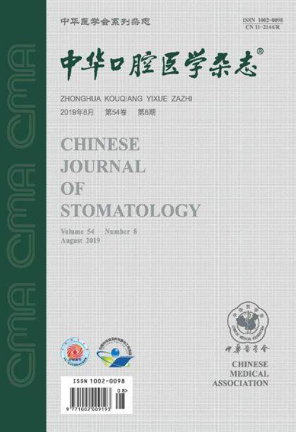
Founded in August 1953, Chinese Journal of Stomatology is a monthly academic journal of stomatology published publicly at home and abroad, sponsored by the Chinese Medical Association and co-sponsored by the Chinese Stomatology Association. It mainly reports the leading scientific research results and clinical diagnosis and treatment experience in the field of oral medicine, as well as the basic theoretical research that has a guiding role in oral clinical practice and is closely combined with oral clinical practice.
Chinese Journal of Over the years, Stomatology has been published in Medline, Scopus database, Toxicology Abstracts Database, Chemical Abstracts Database, American Cancer database, Russian Abstracts database, China Core Journal of Science and Technology, Peking University Core Journal, CSCD and other more than 20 important journals at home and abroad Physical medicine database and retrieval system included.

 求助内容:
 应助结果提醒方式:


 京公网安备 11010802042870号


