{"title":"Histochemical staining optimization of a sperm chromatin dispersion test for the evaluation of DNA fragmentation in bovine spermatozoa.","authors":"Vasiliki Sapanidou, Konstantinos Petropoulos, Dimitrios Kalogiannis, Christos Balaskas","doi":"10.1080/10520295.2026.2737877","DOIUrl":null,"url":null,"abstract":"<p><p>Sperm DNA integrity is a crucial determinant of fertility and early embryonic development. The Sperm Chromatin Dispersion (SCD) test provides a simple, practical, and cost-effective tool for assessing DNA fragmentation; however, the diagnostic reliability depends heavily on the staining technique used to visualize halos of dispersed chromatin. This study evaluated and optimized several commonly and widely used histochemical stains associated with a modified SCD protocol in bovine spermatozoa to improve accuracy and interpretation; hence providing new and useful data for scientists in the field. Frozen-thawed semen samples from five fertile bulls were processed using SCD procedures and stained with Toluidine Blue (TB), Giemsa (G), May Grünwald-Giemsa (MGG), Eosin Thiazine (ET), Feulgen & Rossenbeck (F&R), Hematoxylin & Eosin (H&E), Nuclear Fast Red (NFR), Eosin-Nigrosine (EN), and various modifications of the above stains. Most stains and their modifications (TB, G, ET, NFR, EN) failed to provide consistent or distinguishable halos, whilst specific modifications of MGG, F&R, and H&E produced reproducible halos. The MGG-modified protocol enabled halo visualization but introduced notable nonspecific background staining that reduced distinctiveness of halos. Specific modified protocols of F&R and H&E also visualized halos but reacted variably with agarose: F&R did not produce nonspecific background staining while H&E did. In conclusion, our results demonstrate that specific modifications of MGG, F&R, and H&E visualized halos and thus are suitable for SCD test.</p>","PeriodicalId":8970,"journal":{"name":"Biotechnic & Histochemistry","volume":" ","pages":"1-14"},"PeriodicalIF":1.4000,"publicationDate":"2026-09-30","publicationTypes":"Journal Article","fieldsOfStudy":null,"isOpenAccess":false,"openAccessPdf":"","citationCount":"0","resultStr":null,"platform":"Semanticscholar","paperid":null,"PeriodicalName":"Biotechnic & Histochemistry","FirstCategoryId":"5","ListUrlMain":"https://doi.org/10.1080/10520295.2026.2737877","RegionNum":4,"RegionCategory":"生物学","ArticlePicture":[],"TitleCN":null,"AbstractTextCN":null,"PMCID":null,"EPubDate":"","PubModel":"","JCR":"Q4","JCRName":"BIOTECHNOLOGY & APPLIED MICROBIOLOGY","Score":null,"Total":0}
引用次数: 0
Abstract
Sperm DNA integrity is a crucial determinant of fertility and early embryonic development. The Sperm Chromatin Dispersion (SCD) test provides a simple, practical, and cost-effective tool for assessing DNA fragmentation; however, the diagnostic reliability depends heavily on the staining technique used to visualize halos of dispersed chromatin. This study evaluated and optimized several commonly and widely used histochemical stains associated with a modified SCD protocol in bovine spermatozoa to improve accuracy and interpretation; hence providing new and useful data for scientists in the field. Frozen-thawed semen samples from five fertile bulls were processed using SCD procedures and stained with Toluidine Blue (TB), Giemsa (G), May Grünwald-Giemsa (MGG), Eosin Thiazine (ET), Feulgen & Rossenbeck (F&R), Hematoxylin & Eosin (H&E), Nuclear Fast Red (NFR), Eosin-Nigrosine (EN), and various modifications of the above stains. Most stains and their modifications (TB, G, ET, NFR, EN) failed to provide consistent or distinguishable halos, whilst specific modifications of MGG, F&R, and H&E produced reproducible halos. The MGG-modified protocol enabled halo visualization but introduced notable nonspecific background staining that reduced distinctiveness of halos. Specific modified protocols of F&R and H&E also visualized halos but reacted variably with agarose: F&R did not produce nonspecific background staining while H&E did. In conclusion, our results demonstrate that specific modifications of MGG, F&R, and H&E visualized halos and thus are suitable for SCD test.
期刊介绍:
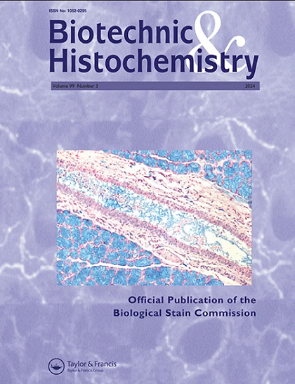
Biotechnic & Histochemistry (formerly Stain technology) is the
official publication of the Biological Stain Commission. The journal has been in continuous publication since 1926.
Biotechnic & Histochemistry is an interdisciplinary journal that embraces all aspects of techniques for visualizing biological processes and entities in cells, tissues and organisms; papers that describe experimental work that employs such investigative methods are appropriate for publication as well.
Papers concerning topics as diverse as applications of histochemistry, immunohistochemistry, in situ hybridization, cytochemical probes, autoradiography, light and electron microscopy, tissue culture, in vivo and in vitro studies, image analysis, cytogenetics, automation or computerization of investigative procedures and other investigative approaches are appropriate for publication regardless of their length. Letters to the Editor and review articles concerning topics of special and current interest also are welcome.

 求助内容:
 应助结果提醒方式:


 京公网安备 11010802042870号


