Emily R. Sekera, Nathaniel Twarog, Rebekah DeVries, P. Jake Slavish, John J. Bowling, Anang A. Shelat
{"title":"SpotOn: A Tool to Enhance Echo Acoustic Standard Deposition for Quantitative Mass Spectrometry Imaging","authors":"Emily R. Sekera, Nathaniel Twarog, Rebekah DeVries, P. Jake Slavish, John J. Bowling, Anang A. Shelat","doi":"10.1021/cbmi.6c00010","DOIUrl":null,"url":null,"abstract":"<p>The quantitative evaluation of therapeutics in the drug\r\ndiscovery\r\npipeline is essential for elucidating their pharmacokinetic and pharmacodynamic\r\nprofiles within complex biological systems. Traditionally, autoradiography\r\nand liquid chromatography-mass spectrometry (LC-MS) have been employed\r\nfor such analyses. However, matrix-assisted laser desorption ionization\r\nmass spectrometry imaging (MALDI-MSI) is emerging as a promising alternative,\r\noffering a more rapid untargeted workflow. Quantitative MALDI-MSI\r\n(qMSI) relies on accurate calibration to achieve robust and reproducible\r\nquantification of analytes. Here, we present an open-source software\r\ntool, SpotOn, designed to facilitate the discrete deposition of low-volume\r\ncalibration standards onto glass slides or tissue sections. By enabling\r\nprecise placement and significantly reducing the volume of standards\r\nrequired, our tool affords the generation of more calibration points\r\nacross a wider dynamic range, thereby improving the robustness and\r\naccuracy of quantitative analyses. Software was validated using JQ1\r\non both glass slides and murine liver tissue. The resulting calibration\r\ncurves demonstrated strong linearity, with coefficients of determination\r\n(<em>R</em><sup>2</sup>) exceeding 0.92. This tool streamlines\r\nthe qMSI workflow, conserves valuable reagents, and enhances analytical\r\nreliability, making it a helpful resource.</p>","PeriodicalId":53181,"journal":{"name":"Chemical & Biomedical Imaging","volume":"4 9","pages":"2119–2125"},"PeriodicalIF":6.7000,"publicationDate":"2026-09-28","publicationTypes":"Journal Article","fieldsOfStudy":null,"isOpenAccess":false,"openAccessPdf":"","citationCount":"0","resultStr":null,"platform":"Semanticscholar","paperid":null,"PeriodicalName":"Chemical & Biomedical Imaging","FirstCategoryId":"1085","ListUrlMain":"https://pubs.acs.org/doi/10.1021/cbmi.6c00010","RegionNum":0,"RegionCategory":null,"ArticlePicture":[],"TitleCN":null,"AbstractTextCN":null,"PMCID":null,"EPubDate":"2026/7/22 0:00:00","PubModel":"Epub","JCR":"","JCRName":"","Score":null,"Total":0}
引用次数: 0
Abstract
The quantitative evaluation of therapeutics in the drug
discovery
pipeline is essential for elucidating their pharmacokinetic and pharmacodynamic
profiles within complex biological systems. Traditionally, autoradiography
and liquid chromatography-mass spectrometry (LC-MS) have been employed
for such analyses. However, matrix-assisted laser desorption ionization
mass spectrometry imaging (MALDI-MSI) is emerging as a promising alternative,
offering a more rapid untargeted workflow. Quantitative MALDI-MSI
(qMSI) relies on accurate calibration to achieve robust and reproducible
quantification of analytes. Here, we present an open-source software
tool, SpotOn, designed to facilitate the discrete deposition of low-volume
calibration standards onto glass slides or tissue sections. By enabling
precise placement and significantly reducing the volume of standards
required, our tool affords the generation of more calibration points
across a wider dynamic range, thereby improving the robustness and
accuracy of quantitative analyses. Software was validated using JQ1
on both glass slides and murine liver tissue. The resulting calibration
curves demonstrated strong linearity, with coefficients of determination
(R2) exceeding 0.92. This tool streamlines
the qMSI workflow, conserves valuable reagents, and enhances analytical
reliability, making it a helpful resource.
期刊介绍:
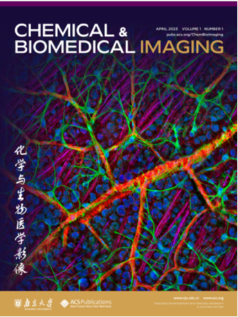
Chemical & Biomedical Imaging is a peer-reviewed open access journal devoted to the publication of cutting-edge research papers on all aspects of chemical and biomedical imaging. This interdisciplinary field sits at the intersection of chemistry physics biology materials engineering and medicine. The journal aims to bring together researchers from across these disciplines to address cutting-edge challenges of fundamental research and applications.Topics of particular interest include but are not limited to:Imaging of processes and reactionsImaging of nanoscale microscale and mesoscale materialsImaging of biological interactions and interfacesSingle-molecule and cellular imagingWhole-organ and whole-body imagingMolecular imaging probes and contrast agentsBioluminescence chemiluminescence and electrochemiluminescence imagingNanophotonics and imagingChemical tools for new imaging modalitiesChemical and imaging techniques in diagnosis and therapyImaging-guided drug deliveryAI and machine learning assisted imaging

 求助内容:
 应助结果提醒方式:


 京公网安备 11010802042870号


