Wenzuo Shang, Jingyu Cui, Xueling Bai, Le Cao, Mangmang Xu, Shuai Jiang, Bo Wu
{"title":"HMGB1 induces mitochondrial dysfunction through the TLR4 and RAGE pathways to promote astrocyte autophagy and PANoptosis in intracerebral hemorrhage.","authors":"Wenzuo Shang, Jingyu Cui, Xueling Bai, Le Cao, Mangmang Xu, Shuai Jiang, Bo Wu","doi":"10.1097/CM9.0000000000004250","DOIUrl":null,"url":null,"abstract":"<p><strong>Background: </strong>Secondary injury is an important factor that leads to a poor prognosis of intracerebral hemorrhage (ICH). Programmed cell death (PCD) plays an important role in secondary injury. After ICH, hyperactivated astrocytes transition from a protective role to a proinflammatory state, releasing neurotoxic factors that amplify neuroinflammation and exacerbate neurological deficits. Through multi-omics sequencing, we found High-mobility group box 1 (HMGB1)may be a key factor . In this study, we investigated its regulatory role in ICH and different PCDs.</p><p><strong>Methods: </strong>The ICH model was established by collagenase injection in vivo and hemoglobin treatment in vitro. Prognosis was assessed using the rotarod test and magnetic resonance imaging (MRI). Cell proliferation activity and growth status were detected using the cell counting kit 8 (CCK-8). Western blotting, enzyme-linked immunosorbent assay (ELISA), and immunofluorescence (IF) were used to detect the related proteins. Transmission electron microscopy (TEM) was used to observe pathological changes. The transferase dUTP nick end labelling (TUNEL) staining and Annexin V/Propidium Iodide (PI) assay were used to detect the apoptosis. Multi-omics experiments were conducted to identify potential pathways of HMGB1 action.</p><p><strong>Results: </strong>The results showed that after ICH, HMGB1 translocated from the nucleus to the cytoplasm and extracellular space. Extracellular HMGB1 upregulated the expression of related molecules, increased the corresponding pathological changes. Multi-omics sequencing suggested that mitochondrial function-related pathways were enriched after ICH. Results showed that increased extracellular HMGB1 aggravated mitochondrial morphology and functional damage and the expression and release of inflammatory factors. Inhibiting TLR4 and RAGE pathways alleviated the HMGB1-mediated injuries.</p><p><strong>Conclusion: </strong>In the acute phase of ICH, HMGB1 promoted poor prognosis by mediating mitochondrial dysfunction through the TLR4 and RAGE pathways, thereby promoting autophagy and PANoptosis while increasing neuroinflammation after ICH.</p>","PeriodicalId":10183,"journal":{"name":"Chinese Medical Journal","volume":" ","pages":""},"PeriodicalIF":9.1000,"publicationDate":"2026-09-21","publicationTypes":"Journal Article","fieldsOfStudy":null,"isOpenAccess":false,"openAccessPdf":"","citationCount":"0","resultStr":null,"platform":"Semanticscholar","paperid":null,"PeriodicalName":"Chinese Medical Journal","FirstCategoryId":"3","ListUrlMain":"https://doi.org/10.1097/CM9.0000000000004250","RegionNum":3,"RegionCategory":"医学","ArticlePicture":[],"TitleCN":null,"AbstractTextCN":null,"PMCID":null,"EPubDate":"","PubModel":"","JCR":"Q1","JCRName":"MEDICINE, GENERAL & INTERNAL","Score":null,"Total":0}
引用次数: 0
Abstract
Background: Secondary injury is an important factor that leads to a poor prognosis of intracerebral hemorrhage (ICH). Programmed cell death (PCD) plays an important role in secondary injury. After ICH, hyperactivated astrocytes transition from a protective role to a proinflammatory state, releasing neurotoxic factors that amplify neuroinflammation and exacerbate neurological deficits. Through multi-omics sequencing, we found High-mobility group box 1 (HMGB1)may be a key factor . In this study, we investigated its regulatory role in ICH and different PCDs.
Methods: The ICH model was established by collagenase injection in vivo and hemoglobin treatment in vitro. Prognosis was assessed using the rotarod test and magnetic resonance imaging (MRI). Cell proliferation activity and growth status were detected using the cell counting kit 8 (CCK-8). Western blotting, enzyme-linked immunosorbent assay (ELISA), and immunofluorescence (IF) were used to detect the related proteins. Transmission electron microscopy (TEM) was used to observe pathological changes. The transferase dUTP nick end labelling (TUNEL) staining and Annexin V/Propidium Iodide (PI) assay were used to detect the apoptosis. Multi-omics experiments were conducted to identify potential pathways of HMGB1 action.
Results: The results showed that after ICH, HMGB1 translocated from the nucleus to the cytoplasm and extracellular space. Extracellular HMGB1 upregulated the expression of related molecules, increased the corresponding pathological changes. Multi-omics sequencing suggested that mitochondrial function-related pathways were enriched after ICH. Results showed that increased extracellular HMGB1 aggravated mitochondrial morphology and functional damage and the expression and release of inflammatory factors. Inhibiting TLR4 and RAGE pathways alleviated the HMGB1-mediated injuries.
Conclusion: In the acute phase of ICH, HMGB1 promoted poor prognosis by mediating mitochondrial dysfunction through the TLR4 and RAGE pathways, thereby promoting autophagy and PANoptosis while increasing neuroinflammation after ICH.
期刊介绍:
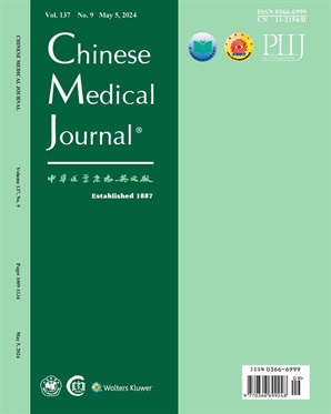
The Chinese Medical Journal (CMJ) is published semimonthly in English by the Chinese Medical Association, and is a peer reviewed general medical journal for all doctors, researchers, and health workers regardless of their medical specialty or type of employment. Established in 1887, it is the oldest medical periodical in China and is distributed worldwide. The journal functions as a window into China’s medical sciences and reflects the advances and progress in China’s medical sciences and technology. It serves the objective of international academic exchange. The journal includes Original Articles, Editorial, Review Articles, Medical Progress, Brief Reports, Case Reports, Viewpoint, Clinical Exchange, Letter,and News,etc. CMJ is abstracted or indexed in many databases including Biological Abstracts, Chemical Abstracts, Index Medicus/Medline, Science Citation Index (SCI), Current Contents, Cancerlit, Health Plan & Administration, Embase, Social Scisearch, Aidsline, Toxline, Biocommercial Abstracts, Arts and Humanities Search, Nuclear Science Abstracts, Water Resources Abstracts, Cab Abstracts, Occupation Safety & Health, etc. In 2007, the impact factor of the journal by SCI is 0.636, and the total citation is 2315.

 求助内容:
 应助结果提醒方式:


 京公网安备 11010802042870号


