
Evanescent-Field Microscopy Combining Resonant Enhancement with Extended Penetration Depth

IF 6 1区 物理与天体物理 Q1 MATERIALS SCIENCE, MULTIDISCIPLINARY
Qihao Sun,Yang Zhao,Zongyan Zhang,Chunzheng Bai,Mingyi Tao,Lei Wu,Zhuyuan Wang,Jiayu Zhang
{"title":"Evanescent-Field\nMicroscopy Combining Resonant Enhancement\nwith Extended Penetration Depth","authors":"Qihao Sun,Yang Zhao,Zongyan Zhang,Chunzheng Bai,Mingyi Tao,Lei Wu,Zhuyuan Wang,Jiayu Zhang","doi":"10.1021/acsphotonics.6c01842","DOIUrl":null,"url":null,"abstract":"Evanescent-field imaging enables selective probing of near-interface regions and has been widely used to visualize cell membrane dynamics, cell–substrate interfaces, nanoparticles, and surface-associated biological structures. Although conventional plasmonic illumination enables sensitive detection of subtle changes in the local dielectric environment through resonant field enhancement, its effective probing range is typically confined to within 100–200 nm of the interface. Here, we present long-range plasmonic illumination microscopy (LRPIM) based on a metal–dielectric multilayer chip. At the sample-facing surface, the chip generates an enhanced evanescent field that penetrates deeply into the sample while preserving low-background near-field excitation. Using individual microspheres as scattering probes, we established a calibration relationship between the evanescent-field penetration depth and the full width at half-maximum (FWHM) of the focused hotspot formed at the exit surface of each microsphere. Based on this calibration, the chip exhibited an electric-field penetration depth approximately 4.3 times that obtained with plasmonic scattering microscopy (PSM), enabling near-surface illumination over a micrometer-scale axial range. Furthermore, LRPIM supports fluorescence imaging, enhancing fluorescence signals from propidium iodide (PI)-stained cells and revealing localized nucleic acid enrichment within their nuclei. We anticipate that LRPIM will provide a straightforward chip-based approach to evanescent-field scattering and fluorescence microscopy, enabling structures to be probed over a micrometer-scale axial range near interfaces.","PeriodicalId":23,"journal":{"name":"ACS Photonics","volume":"30 1","pages":""},"PeriodicalIF":6.0000,"publicationDate":"2026-09-06","publicationTypes":"Journal Article","fieldsOfStudy":null,"isOpenAccess":false,"openAccessPdf":"","citationCount":"0","resultStr":null,"platform":"Semanticscholar","paperid":null,"PeriodicalName":"ACS Photonics","FirstCategoryId":"101","ListUrlMain":"https://doi.org/10.1021/acsphotonics.6c01842","RegionNum":1,"RegionCategory":"物理与天体物理","ArticlePicture":[],"TitleCN":null,"AbstractTextCN":null,"PMCID":null,"EPubDate":"","PubModel":"","JCR":"Q1","JCRName":"MATERIALS SCIENCE, MULTIDISCIPLINARY","Score":null,"Total":0}
引用次数: 0

Abstract

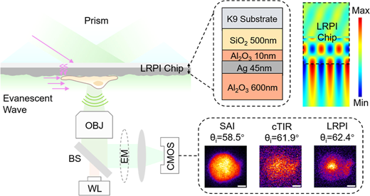
Evanescent-field imaging enables selective probing of near-interface regions and has been widely used to visualize cell membrane dynamics, cell–substrate interfaces, nanoparticles, and surface-associated biological structures. Although conventional plasmonic illumination enables sensitive detection of subtle changes in the local dielectric environment through resonant field enhancement, its effective probing range is typically confined to within 100–200 nm of the interface. Here, we present long-range plasmonic illumination microscopy (LRPIM) based on a metal–dielectric multilayer chip. At the sample-facing surface, the chip generates an enhanced evanescent field that penetrates deeply into the sample while preserving low-background near-field excitation. Using individual microspheres as scattering probes, we established a calibration relationship between the evanescent-field penetration depth and the full width at half-maximum (FWHM) of the focused hotspot formed at the exit surface of each microsphere. Based on this calibration, the chip exhibited an electric-field penetration depth approximately 4.3 times that obtained with plasmonic scattering microscopy (PSM), enabling near-surface illumination over a micrometer-scale axial range. Furthermore, LRPIM supports fluorescence imaging, enhancing fluorescence signals from propidium iodide (PI)-stained cells and revealing localized nucleic acid enrichment within their nuclei. We anticipate that LRPIM will provide a straightforward chip-based approach to evanescent-field scattering and fluorescence microscopy, enabling structures to be probed over a micrometer-scale axial range near interfaces.
Abstract Image

结合共振增强和扩展穿透深度的倏逝场显微镜
倏逝场成像能够选择性探测近界面区域,并已广泛用于可视化细胞膜动力学、细胞-底物界面、纳米颗粒和表面相关生物结构。虽然传统的等离子体照明可以通过共振场增强来灵敏地探测局部介质环境的细微变化,但其有效探测范围通常限制在界面的100 - 200nm范围内。在这里,我们提出了基于金属介电介质多层芯片的远程等离子体照明显微镜(LRPIM)。在样品表面,芯片产生一个增强的倏逝场,该场深入样品,同时保持低背景近场激发。利用单个微球作为散射探针,建立了消弱场穿透深度与每个微球出口表面形成的聚焦热点的半最大值全宽度之间的标定关系。基于此校准,该芯片显示出的电场穿透深度约为等离子体散射显微镜(PSM)的4.3倍,可以在微米尺度的轴向范围内实现近表面照明。此外,LRPIM支持荧光成像,增强碘化丙啶(PI)染色细胞的荧光信号,并显示其细胞核内的局部核酸富集。我们预计LRPIM将提供一种直接的基于芯片的方法来进行倏逝场散射和荧光显微镜,使结构能够在靠近界面的微米尺度轴向范围内进行探测。
本文章由计算机程序翻译,如有差异,请以英文原文为准。
求助全文
约1分钟内获得全文 求助全文
来源期刊
ACS Photonics
ACS Photonics NANOSCIENCE & NANOTECHNOLOGY-MATERIALS SCIENCE, MULTIDISCIPLINARY
CiteScore
11.90
自引率
5.70%
发文量
438
审稿时长
2.3 months
期刊介绍: Published as soon as accepted and summarized in monthly issues, ACS Photonics will publish Research Articles, Letters, Perspectives, and Reviews, to encompass the full scope of published research in this field.

×
引用
GB/T 7714-2015
复制
MLA
复制
APA
复制
导出至
BibTeX EndNote RefMan NoteFirst NoteExpress
×
提示
您的信息不完整,为了账户安全,请先补充。
现在去补充
×
提示
您因"违规操作"
具体请查看互助需知
我知道了
×
提示
确定
请完成安全验证×
copy
已复制链接
快去分享给好友吧!
我知道了
右上角分享
点击右上角分享
0
联系我们:info@booksci.cn Book学术提供免费学术资源搜索服务,方便国内外学者检索中英文文献。致力于提供最便捷和优质的服务体验。 Copyright © 2023 布克学术 All rights reserved.
京ICP备2023020795号-1
ghs 京公网安备 11010802042870号
Book学术文献互助
Book学术文献互助群
群 号:604180095
Book学术官方微信
小红书


