
Single-Step Expansion SMLM Enables Molecular-Resolution Imaging in Cells and Isolated Proteins
IF 6.7
引用次数: 0
Abstract
Expansion microscopy (ExM) enables nanoscale fluorescence imaging on standard microscopes, but its combination with single-molecule localization microscopy (SMLM) remains challenging due to the incompatibility of expanded hydrogels with photoswitching buffers. Here, we introduce a single-step expansion microscopy approach that enables SMLM using spontaneously blinking dyes in 6–14× expanded samples, without re-embedding or buffer exchange. Using this approach, we achieve nanometer-scale spatial resolution by resolving the organization of the nuclear pore complex and the molecular structure of recombinant homotrimeric proliferating cell nuclear antigen.
单步扩展SMLM实现细胞和分离蛋白的分子分辨率成像。
膨胀显微镜(ExM)可以在标准显微镜上进行纳米级荧光成像,但由于膨胀水凝胶与光电开关缓冲液的不相容性,它与单分子定位显微镜(SMLM)的结合仍然具有挑战性。在这里,我们介绍了一种单步扩展显微镜方法,使SMLM在6-14倍扩展的样品中使用自发闪烁的染料,而不需要重新嵌入或交换缓冲液。利用这种方法,我们通过解析核孔复合物的组织和重组同源三聚体增殖细胞核抗原的分子结构,实现了纳米尺度的空间分辨率。
本文章由计算机程序翻译,如有差异,请以英文原文为准。
求助全文
约1分钟内获得全文
求助全文
来源期刊
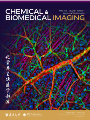
Chemical & Biomedical Imaging
化学与生物成像-
CiteScore
1.00
自引率
0.00%
发文量
0
期刊介绍:
Chemical & Biomedical Imaging is a peer-reviewed open access journal devoted to the publication of cutting-edge research papers on all aspects of chemical and biomedical imaging. This interdisciplinary field sits at the intersection of chemistry physics biology materials engineering and medicine. The journal aims to bring together researchers from across these disciplines to address cutting-edge challenges of fundamental research and applications.Topics of particular interest include but are not limited to:Imaging of processes and reactionsImaging of nanoscale microscale and mesoscale materialsImaging of biological interactions and interfacesSingle-molecule and cellular imagingWhole-organ and whole-body imagingMolecular imaging probes and contrast agentsBioluminescence chemiluminescence and electrochemiluminescence imagingNanophotonics and imagingChemical tools for new imaging modalitiesChemical and imaging techniques in diagnosis and therapyImaging-guided drug deliveryAI and machine learning assisted imaging
 求助内容:
 应助结果提醒方式:


 京公网安备 11010802042870号


