Alvin Karaqi, Philipp Braken, Jana Nelson, Wolf Petersen, Sergio Canuto, Richard Glaab
{"title":"Harvesting the tendon of the rectus femoris muscle as a graft for reconstructive ligament surgery on the knee joint.","authors":"Alvin Karaqi, Philipp Braken, Jana Nelson, Wolf Petersen, Sergio Canuto, Richard Glaab","doi":"10.1007/s00064-026-00947-8","DOIUrl":null,"url":null,"abstract":"<p><strong>Objective: </strong>Harvesting a strip from the distal rectus femoris tendon as an autograft for ligament reconstruction of the knee or other joints.</p><p><strong>Indications: </strong>Ligament reconstructions of the knee, both primary and revision procedures.</p><p><strong>Contraindications: </strong>Relative: athletes in jumping sports requiring rapid recovery of explosive strength.</p><p><strong>Surgical technique: </strong>Palpation of the distal quadriceps tendon at the \"fusion zone,\" approximately 5 cm proximal to the superior patellar pole. A 3-4 cm longitudinal skin incision is made at the junction of the lateral and middle third or centrally. The quadriceps tendon is exposed and identified proximally. After identification, two parallel incisions create an 8-10 mm wide graft. The tendon strip is mobilized with a clamp just above the fusion zone and separated from the deeper layers. Distal detachment of the graft can be performed either (a) with a scalpel after whipstitching of the free end or (b) with a closed tendon stripper following proximal release under tension. The preparation is then extended proximally by 7-8 cm with scissors and bluntly separated from deeper layers with the index finger. An open or closed tendon stripper (8-9 mm) is advanced proximally with the knee in 20° flexion until complete graft harvest. Rotational movements should be avoided.</p><p><strong>Postoperative management: </strong>Rehabilitation follows the protocol of the corresponding ligament reconstruction. No specific measures are required for the donor site.</p><p><strong>Results: </strong>Radiological assessment demonstrated a mean distal rectus femoris tendon length of 39 cm (32-47 cm). In cadaveric studies, the technique was feasible and reproducible. Clinically, the graft was used in 103 patients, with a mean graft diameter of 8.3 mm. In a few cases, hamstring augmentation was required. Complications such as arthrofibrosis or donor-site hematoma were rare and successfully treated. External studies confirmed the suitability of the rectus femoris tendon, particularly for revision anterior cruciate ligament reconstruction.</p>","PeriodicalId":54677,"journal":{"name":"Operative Orthopadie Und Traumatologie","volume":" ","pages":""},"PeriodicalIF":1.1000,"publicationDate":"2026-06-23","publicationTypes":"Journal Article","fieldsOfStudy":null,"isOpenAccess":false,"openAccessPdf":"","citationCount":"0","resultStr":null,"platform":"Semanticscholar","paperid":null,"PeriodicalName":"Operative Orthopadie Und Traumatologie","FirstCategoryId":"3","ListUrlMain":"https://doi.org/10.1007/s00064-026-00947-8","RegionNum":4,"RegionCategory":"医学","ArticlePicture":[],"TitleCN":null,"AbstractTextCN":null,"PMCID":null,"EPubDate":"","PubModel":"","JCR":"Q3","JCRName":"ORTHOPEDICS","Score":null,"Total":0}
引用次数: 0
Abstract
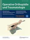
Objective: Harvesting a strip from the distal rectus femoris tendon as an autograft for ligament reconstruction of the knee or other joints.
Indications: Ligament reconstructions of the knee, both primary and revision procedures.
Contraindications: Relative: athletes in jumping sports requiring rapid recovery of explosive strength.
Surgical technique: Palpation of the distal quadriceps tendon at the "fusion zone," approximately 5 cm proximal to the superior patellar pole. A 3-4 cm longitudinal skin incision is made at the junction of the lateral and middle third or centrally. The quadriceps tendon is exposed and identified proximally. After identification, two parallel incisions create an 8-10 mm wide graft. The tendon strip is mobilized with a clamp just above the fusion zone and separated from the deeper layers. Distal detachment of the graft can be performed either (a) with a scalpel after whipstitching of the free end or (b) with a closed tendon stripper following proximal release under tension. The preparation is then extended proximally by 7-8 cm with scissors and bluntly separated from deeper layers with the index finger. An open or closed tendon stripper (8-9 mm) is advanced proximally with the knee in 20° flexion until complete graft harvest. Rotational movements should be avoided.
Postoperative management: Rehabilitation follows the protocol of the corresponding ligament reconstruction. No specific measures are required for the donor site.
Results: Radiological assessment demonstrated a mean distal rectus femoris tendon length of 39 cm (32-47 cm). In cadaveric studies, the technique was feasible and reproducible. Clinically, the graft was used in 103 patients, with a mean graft diameter of 8.3 mm. In a few cases, hamstring augmentation was required. Complications such as arthrofibrosis or donor-site hematoma were rare and successfully treated. External studies confirmed the suitability of the rectus femoris tendon, particularly for revision anterior cruciate ligament reconstruction.
期刊介绍:
Orthopedics and Traumatology is directed toward all orthopedic surgeons, trauma-tologists, hand surgeons, specialists in sports injuries, orthopedics and rheumatology as well as gene-al surgeons who require access to reliable information on current operative methods to ensure the quality of patient advice, preoperative planning, and postoperative care.
The journal presents established and new operative procedures in uniformly structured and extensively illustrated contributions. All aspects are presented step-by-step from indications, contraindications, patient education, and preparation of the operation right through to postoperative care. The advantages and disadvantages, possible complications, deficiencies and risks of the methods as well as significant results with their evaluation criteria are discussed. To allow the reader to assess the outcome, results are detailed and based on internationally recognized scoring systems.
Orthopedics and Traumatology facilitates effective advancement and further education for all those active in both special and conservative fields of orthopedics, traumatology, and general surgery, offers sup-port for therapeutic decision-making, and provides – more than 30 years after its first publication – constantly expanding and up-to-date teaching on operative techniques.

 求助内容:
 应助结果提醒方式:


 京公网安备 11010802042870号


