Y Liang, Y M Qiu, Z F Liu, J He, Z Zhou, L Q Yan, X Lu, Y Yang, H Wang, Z Z Wu, Z X Ning, W H Xia
{"title":"[PRDX1 attenuates hypertensive endothelial dysfunction by inhibiting mTOR/p70S6K signaling].","authors":"Y Liang, Y M Qiu, Z F Liu, J He, Z Zhou, L Q Yan, X Lu, Y Yang, H Wang, Z Z Wu, Z X Ning, W H Xia","doi":"10.3760/cma.j.cn112148-20250918-00662","DOIUrl":null,"url":null,"abstract":"<p><p><b>Objective:</b> To explore the role and molecular mechanism of peroxiredoxin 1 (PRDX1) in hypertension-induced endothelial dysfunction. <b>Methods:</b> (1) Bioinformatics analysis: A total of 40 C57BL/6J mice aged 8-10 weeks (20-25 g) were randomly divided into the saline group and angiotensin Ⅱ (AngⅡ, 0.8 mg·kg⁻¹·d⁻¹) group, with 20 mice in each group. After 4 consecutive weeks of intervention, mice were sacrificed, and thoracic aortic tissues were collected for transcriptome sequencing. Gene Ontology functional annotation and Kyoto Encyclopedia of Genes and Genomes pathway enrichment analysis were performed on differentially expressed genes. (2) Cell experiments: Human umbilical vein endothelial cells (HUVECs) were divided into the control group (endothelial cell culture medium) and the AngⅡ intervention group (medium containing 10<sup>-</sup>⁶ mol/L AngⅡ). Wound healing assay, cell adhesion assay, and Transwell assay were used to assess cell migration and adhesion. Lentiviral or small interfering RNA (siRNA) transfection was performed to achieve PRDX1 overexpression and knockdown, respectively. The overexpression experiment was divided into the LV-NC (negative control lentivirus) group, Ang Ⅱ+LV-NC group, LV-PRDX1 (PRDX1 overexpression lentivirus) group and Ang Ⅱ+LV-PRDX1 group. The knockdown experiment was divided into the NC-siRNA (negative control siRNA) group, si-PRDX1 group, NC-siRNA+rapamycin (50 nmol/L) group and si-PRDX1+rapamycin group. Immunofluorescence staining was applied to detect intracellular reactive oxygen species level. Quantitative reverse transcription-polymerase chain reaction was used to detect the mRNA expression levels of PRDX1 and mammalian target of rapamycin (mTOR). Western blot was adopted to determine the total protein and phosphorylation levels of PRDX1, mTOR, p70 ribosomal S6 kinase (p70S6K) 1 and endothelial nitric oxide synthase (eNOS). Co-immunoprecipitation assay was used to verify the protein interaction between PRDX1 and mTOR. Nitrate reductase method was used to measure cellular nitric oxide (NO) content. (3) Animal experiments: Forty C57BL/6J mice aged 8-10 weeks (20-25 g) were used to construct the PRDX1 overexpression model via adeno-associated virus serotype 9 (AAV9) vector. Mice were assigned into 4 groups with 10 animals per group: saline+AAV9-GFP (empty vector) group, saline+AAV9-PRDX1 (recombinant virus) group, AngⅡ+AAV9-GFP group, and AngⅡ+AAV9-PRDX1 group. Systolic blood pressure and diastolic blood pressure of mice in each group were dynamically monitored at day 0, 7, 14, 21 and 28 after modeling. Plasma NO level was detected by the nitrate reductase method. After sacrifice, isolated thoracic aortic tissues were subjected to morphological and pathological staining analysis, and a microvascular tension measurement system was used to evaluate the acetylcholine-mediated endothelium-dependent vasodilation function. <b>Results:</b> (1) Bioinformatics analysis: Transcriptome sequencing revealed that numerous differentially expressed genes were identified in the thoracic aorta of mice in the AngⅡ group compared with the saline group. These genes were mainly enriched in biological processes closely associated with oxidative stress, such as reactive oxygen species metabolism and oxidative phosphorylation regulation. (2) Cell experiments: Compared with the control group, HUVECs in the AngⅡ intervention group presented decreased protein and mRNA levels of PRDX1, as well as elevated phosphorylation levels of mTOR and p70S6K1 (all <i>P</i><0.05). Compared with the LV-NC group, the LV-PRDX1 group showed higher PRDX1 mRNA expression, lower reactive oxygen species levels, enhanced cell migration and adhesion capacities, and increased NO content (all <i>P</i><0.05). In contrast with the AngⅡ+LV-NC group, the AngⅡ+LV-PRDX1 group exhibited reduced phosphorylation levels of mTOR and p70S6K1 and increased eNOS phosphorylation level (all <i>P</i><0.05). In addition, relative to the NC-siRNA group, the si-PRDX1 group had higher reactive oxygen species levels and elevated phosphorylation of mTOR and p70S6K1, accompanied by decreased NO content, reduced eNOS phosphorylation, and weakened cell migration and adhesion abilities (all <i>P</i><0.05). Compared with the si-PRDX1 group, the above abnormal changes were partially reversed in the si-PRDX1+rapamycin group (all <i>P</i><0.05). Co-immunoprecipitation assay confirmed a protein interaction between PRDX1 and mTOR. (3) Animal experiments: In comparison with the saline+AAV9-GFP group, the AngⅡ+AAV9-GFP group had higher systolic and diastolic blood pressure, lower plasma NO level, thicker thoracic aortic media, increased collagen deposition, disordered arrangement of elastic fibers, and impaired endothelium-dependent vasodilation in response to acetylcholine (all <i>P</i><0.05). Notably, the AngⅡ+AAV9-PRDX1 group showed lower systolic and diastolic blood pressure, alleviated pathological damage of the thoracic aorta, improved endothelium-dependent vasodilation function, and higher plasma NO level than the AngⅡ+AAV9-GFP group (all <i>P</i><0.05). <b>Conclusion:</b> PRDX1 can inhibit the excessive activation of the mTOR/p70S6K signaling pathway by scavenging reactive oxygen species and promoting NO production, thereby regulating eNOS activity and ameliorating endothelial dysfunction and vascular injury under hypertensive conditions. Targeted regulation of the PRDX1/ROS/mTOR/p70S6K signaling axis is expected to provide a novel therapeutic target and intervention strategy for the prevention and treatment of hypertensive vascular diseases.</p>","PeriodicalId":38755,"journal":{"name":"中华心血管病杂志","volume":"54 6","pages":"671-684"},"PeriodicalIF":0.0000,"publicationDate":"2026-06-24","publicationTypes":"Journal Article","fieldsOfStudy":null,"isOpenAccess":false,"openAccessPdf":"","citationCount":"0","resultStr":null,"platform":"Semanticscholar","paperid":null,"PeriodicalName":"中华心血管病杂志","FirstCategoryId":"3","ListUrlMain":"https://doi.org/10.3760/cma.j.cn112148-20250918-00662","RegionNum":0,"RegionCategory":null,"ArticlePicture":[],"TitleCN":null,"AbstractTextCN":null,"PMCID":null,"EPubDate":"","PubModel":"","JCR":"Q3","JCRName":"Medicine","Score":null,"Total":0}
引用次数: 0
Abstract
Objective: To explore the role and molecular mechanism of peroxiredoxin 1 (PRDX1) in hypertension-induced endothelial dysfunction. Methods: (1) Bioinformatics analysis: A total of 40 C57BL/6J mice aged 8-10 weeks (20-25 g) were randomly divided into the saline group and angiotensin Ⅱ (AngⅡ, 0.8 mg·kg⁻¹·d⁻¹) group, with 20 mice in each group. After 4 consecutive weeks of intervention, mice were sacrificed, and thoracic aortic tissues were collected for transcriptome sequencing. Gene Ontology functional annotation and Kyoto Encyclopedia of Genes and Genomes pathway enrichment analysis were performed on differentially expressed genes. (2) Cell experiments: Human umbilical vein endothelial cells (HUVECs) were divided into the control group (endothelial cell culture medium) and the AngⅡ intervention group (medium containing 10-⁶ mol/L AngⅡ). Wound healing assay, cell adhesion assay, and Transwell assay were used to assess cell migration and adhesion. Lentiviral or small interfering RNA (siRNA) transfection was performed to achieve PRDX1 overexpression and knockdown, respectively. The overexpression experiment was divided into the LV-NC (negative control lentivirus) group, Ang Ⅱ+LV-NC group, LV-PRDX1 (PRDX1 overexpression lentivirus) group and Ang Ⅱ+LV-PRDX1 group. The knockdown experiment was divided into the NC-siRNA (negative control siRNA) group, si-PRDX1 group, NC-siRNA+rapamycin (50 nmol/L) group and si-PRDX1+rapamycin group. Immunofluorescence staining was applied to detect intracellular reactive oxygen species level. Quantitative reverse transcription-polymerase chain reaction was used to detect the mRNA expression levels of PRDX1 and mammalian target of rapamycin (mTOR). Western blot was adopted to determine the total protein and phosphorylation levels of PRDX1, mTOR, p70 ribosomal S6 kinase (p70S6K) 1 and endothelial nitric oxide synthase (eNOS). Co-immunoprecipitation assay was used to verify the protein interaction between PRDX1 and mTOR. Nitrate reductase method was used to measure cellular nitric oxide (NO) content. (3) Animal experiments: Forty C57BL/6J mice aged 8-10 weeks (20-25 g) were used to construct the PRDX1 overexpression model via adeno-associated virus serotype 9 (AAV9) vector. Mice were assigned into 4 groups with 10 animals per group: saline+AAV9-GFP (empty vector) group, saline+AAV9-PRDX1 (recombinant virus) group, AngⅡ+AAV9-GFP group, and AngⅡ+AAV9-PRDX1 group. Systolic blood pressure and diastolic blood pressure of mice in each group were dynamically monitored at day 0, 7, 14, 21 and 28 after modeling. Plasma NO level was detected by the nitrate reductase method. After sacrifice, isolated thoracic aortic tissues were subjected to morphological and pathological staining analysis, and a microvascular tension measurement system was used to evaluate the acetylcholine-mediated endothelium-dependent vasodilation function. Results: (1) Bioinformatics analysis: Transcriptome sequencing revealed that numerous differentially expressed genes were identified in the thoracic aorta of mice in the AngⅡ group compared with the saline group. These genes were mainly enriched in biological processes closely associated with oxidative stress, such as reactive oxygen species metabolism and oxidative phosphorylation regulation. (2) Cell experiments: Compared with the control group, HUVECs in the AngⅡ intervention group presented decreased protein and mRNA levels of PRDX1, as well as elevated phosphorylation levels of mTOR and p70S6K1 (all P<0.05). Compared with the LV-NC group, the LV-PRDX1 group showed higher PRDX1 mRNA expression, lower reactive oxygen species levels, enhanced cell migration and adhesion capacities, and increased NO content (all P<0.05). In contrast with the AngⅡ+LV-NC group, the AngⅡ+LV-PRDX1 group exhibited reduced phosphorylation levels of mTOR and p70S6K1 and increased eNOS phosphorylation level (all P<0.05). In addition, relative to the NC-siRNA group, the si-PRDX1 group had higher reactive oxygen species levels and elevated phosphorylation of mTOR and p70S6K1, accompanied by decreased NO content, reduced eNOS phosphorylation, and weakened cell migration and adhesion abilities (all P<0.05). Compared with the si-PRDX1 group, the above abnormal changes were partially reversed in the si-PRDX1+rapamycin group (all P<0.05). Co-immunoprecipitation assay confirmed a protein interaction between PRDX1 and mTOR. (3) Animal experiments: In comparison with the saline+AAV9-GFP group, the AngⅡ+AAV9-GFP group had higher systolic and diastolic blood pressure, lower plasma NO level, thicker thoracic aortic media, increased collagen deposition, disordered arrangement of elastic fibers, and impaired endothelium-dependent vasodilation in response to acetylcholine (all P<0.05). Notably, the AngⅡ+AAV9-PRDX1 group showed lower systolic and diastolic blood pressure, alleviated pathological damage of the thoracic aorta, improved endothelium-dependent vasodilation function, and higher plasma NO level than the AngⅡ+AAV9-GFP group (all P<0.05). Conclusion: PRDX1 can inhibit the excessive activation of the mTOR/p70S6K signaling pathway by scavenging reactive oxygen species and promoting NO production, thereby regulating eNOS activity and ameliorating endothelial dysfunction and vascular injury under hypertensive conditions. Targeted regulation of the PRDX1/ROS/mTOR/p70S6K signaling axis is expected to provide a novel therapeutic target and intervention strategy for the prevention and treatment of hypertensive vascular diseases.
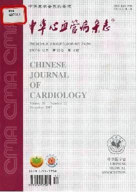
中华心血管病杂志Medicine-Cardiology and Cardiovascular Medicine
CiteScore
1.40
自引率
0.00%
发文量
10577
期刊介绍:
The Chinese Journal of Cardiology , established in February 1973, is one of the major academic medical journals sponsored by the Chinese Medical Association and a leading periodical in the field of cardiology in China. It specializes in cardiology and related disciplines with a readership of more than 25 000. The journal publishes editorials and guidelines as well as important original articles on clinical and experimental investigations, reflecting achievements made in China and promoting academic communication between domestic and foreign cardiologists. The journal includes the following columns: Editorials, Strategies, Comments, Clinical Investigations, Experimental Investigations, Epidemiology and Prevention, Lectures, Comprehensive Reviews, Continuing Medical Education, etc.

 求助内容:
 应助结果提醒方式:


 京公网安备 11010802042870号


