Bevacizumab alleviates kidney damage by modulating inflammation, necroptosis and apoptosis: a preclinical study of renal ischaemia/reperfusion injury in rats
{"title":"Bevacizumab alleviates kidney damage by modulating inflammation, necroptosis and apoptosis: a preclinical study of renal ischaemia/reperfusion injury in rats","authors":"Ali M. Janabi, Heider Qassam, Nadhim K. Hante","doi":"10.1007/s10735-025-10635-9","DOIUrl":null,"url":null,"abstract":"<div><p>Renal ischemia/reperfusion injury is a critical clinical problem caused by kidney and heart surgery and can lead to acute kidney injury (AKI). Bevacizumab is a humanized monoclonal antibody that binds to circulating soluble isoforms of VEGF-A, thereby inhibiting the activation of VEGF molecular pathways and eliciting antiangiogenic effects. This study assessed the nephroprotective potential of bevacizumab in a rat model of renal ischemia/reperfusion injury (I/R). Twenty-four Sprague–Dawley rats were allocated into four groups: Sham, I/R, I/R + normal saline, and I/R + bevacizumab. The sham group was subjected to laparotomy without I/R induction. The I/R, I/R + normal saline, and I/R + bevacizumab groups were subjected to 30 min of bilateral renal ischemia, followed by 24 h of reperfusion. The rats in the I/R + normal saline and I/R + bevacizumab groups were administered normal saline (vehicle for bevacizumab) and 0.1 mg/kg bevacizumab via intraperitoneal injection 60 min before ischemia, respectively. Renal damage markers (creatinine and KIM-1), inflammatory and oxidative markers (TNF-α, IL-1β, NF-κB, F8-isoprostane and SOD), and an apoptotic marker (caspase-3) were measured via ELISA. Nrf2 and MLKL were assessed by IHC, and RIPK1 and HO-1 were assessed by RT‒qPCR, in addition to histological examination and molecular docking. Compared with the sham group, the I/R and I/R + normal saline groups presented significant increases in creatinine, KIM-1, NF-κB, TNF-α, IL-1β, F8-isoprostane, caspase-3, RIPK1, and MLKL and a reduction in SOD. Compared with those in the sham group, the histological findings in the I/R and I/R + normal saline groups revealed notable structural damage. Conversely, bevacizumab significantly reduced renal damage, inflammatory marker levels, cellular death, and histopathological findings. In bevacizumab-treated rats, the nuclear translocation of Nrf2 and HO-1 increased. Moreover, molecular docking analysis revealed that bevacizumab interacted with Keap1. Bevacizumab has nephroprotective effects against renal IRI by diminishing inflammation, necroptosis, apoptosis, and necrosis through the activation of the Nrf2/HO-1 pathway and the inhibition of the RIPK1/MLKL pathway.</p></div>","PeriodicalId":650,"journal":{"name":"Journal of Molecular Histology","volume":"56 6","pages":""},"PeriodicalIF":2.2000,"publicationDate":"2025-10-22","publicationTypes":"Journal Article","fieldsOfStudy":null,"isOpenAccess":false,"openAccessPdf":"","citationCount":"0","resultStr":null,"platform":"Semanticscholar","paperid":null,"PeriodicalName":"Journal of Molecular Histology","FirstCategoryId":"99","ListUrlMain":"https://link.springer.com/article/10.1007/s10735-025-10635-9","RegionNum":4,"RegionCategory":"生物学","ArticlePicture":[],"TitleCN":null,"AbstractTextCN":null,"PMCID":null,"EPubDate":"","PubModel":"","JCR":"Q3","JCRName":"CELL BIOLOGY","Score":null,"Total":0}
引用次数: 0
Abstract
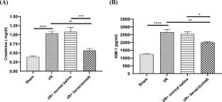
Renal ischemia/reperfusion injury is a critical clinical problem caused by kidney and heart surgery and can lead to acute kidney injury (AKI). Bevacizumab is a humanized monoclonal antibody that binds to circulating soluble isoforms of VEGF-A, thereby inhibiting the activation of VEGF molecular pathways and eliciting antiangiogenic effects. This study assessed the nephroprotective potential of bevacizumab in a rat model of renal ischemia/reperfusion injury (I/R). Twenty-four Sprague–Dawley rats were allocated into four groups: Sham, I/R, I/R + normal saline, and I/R + bevacizumab. The sham group was subjected to laparotomy without I/R induction. The I/R, I/R + normal saline, and I/R + bevacizumab groups were subjected to 30 min of bilateral renal ischemia, followed by 24 h of reperfusion. The rats in the I/R + normal saline and I/R + bevacizumab groups were administered normal saline (vehicle for bevacizumab) and 0.1 mg/kg bevacizumab via intraperitoneal injection 60 min before ischemia, respectively. Renal damage markers (creatinine and KIM-1), inflammatory and oxidative markers (TNF-α, IL-1β, NF-κB, F8-isoprostane and SOD), and an apoptotic marker (caspase-3) were measured via ELISA. Nrf2 and MLKL were assessed by IHC, and RIPK1 and HO-1 were assessed by RT‒qPCR, in addition to histological examination and molecular docking. Compared with the sham group, the I/R and I/R + normal saline groups presented significant increases in creatinine, KIM-1, NF-κB, TNF-α, IL-1β, F8-isoprostane, caspase-3, RIPK1, and MLKL and a reduction in SOD. Compared with those in the sham group, the histological findings in the I/R and I/R + normal saline groups revealed notable structural damage. Conversely, bevacizumab significantly reduced renal damage, inflammatory marker levels, cellular death, and histopathological findings. In bevacizumab-treated rats, the nuclear translocation of Nrf2 and HO-1 increased. Moreover, molecular docking analysis revealed that bevacizumab interacted with Keap1. Bevacizumab has nephroprotective effects against renal IRI by diminishing inflammation, necroptosis, apoptosis, and necrosis through the activation of the Nrf2/HO-1 pathway and the inhibition of the RIPK1/MLKL pathway.
期刊介绍:
The Journal of Molecular Histology publishes results of original research on the localization and expression of molecules in animal cells, tissues and organs. Coverage includes studies describing novel cellular or ultrastructural distributions of molecules which provide insight into biochemical or physiological function, development, histologic structure and disease processes.
Major research themes of particular interest include:
- Cell-Cell and Cell-Matrix Interactions;
- Connective Tissues;
- Development and Disease;
- Neuroscience.
Please note that the Journal of Molecular Histology does not consider manuscripts dealing with the application of immunological or other probes on non-standard laboratory animal models unless the results are clearly of significant and general biological importance.
The Journal of Molecular Histology publishes full-length original research papers, review articles, short communications and letters to the editors. All manuscripts are typically reviewed by two independent referees. The Journal of Molecular Histology is a continuation of The Histochemical Journal.

 求助内容:
求助内容: 应助结果提醒方式:


