Jie Yang, Wei Liu, Jie An, Cheng Jiao, Zhi Li, Shuai Qi, Chen Hao, Yao Zhang, Hui-Lin Wang, Jun Guo
{"title":"Case Report: Primary breast leiomyosarcoma in an 84-year-old male.","authors":"Jie Yang, Wei Liu, Jie An, Cheng Jiao, Zhi Li, Shuai Qi, Chen Hao, Yao Zhang, Hui-Lin Wang, Jun Guo","doi":"10.3389/fonc.2025.1660377","DOIUrl":null,"url":null,"abstract":"<p><p>Primary breast leiomyosarcoma is an extremely rare malignancy originating from mesenchymal tissue. Fewer than 10 male cases have been reported globally. This paper reports an 84-year-old male patient. This represents the oldest reported case in the current literature. A painless, slowly enlarging mass was present in his right breast. The mass had a 10-year history. This contrasts sharply with the typically rapidly progressive pattern documented in previous literature. Clinical examination revealed a mobile mass measuring 12 cm × 10 cm in the right breast. No lymphadenopathy was detected. Ultrasound showed a hypoechoic lesion classified as BI-RADS 4a. Magnetic resonance imaging demonstrated plateau-type enhancement. The patient underwent simple mastectomy. Axillary lymph node dissection was not performed. Postoperative pathology and immunohistochemistry confirmed the diagnosis of breast leiomyosarcoma. The patient declined adjuvant radiotherapy. Follow-up at 6 months postoperatively showed no local recurrence or metastasis. This case indicates several points to clinicians. Immunohistochemistry serves as the cornerstone for diagnosing spindle cell tumors of the breast. R0 surgical resection constitutes the core approach for achieving cure. Decisions regarding adjuvant therapy require full consideration of host age and tumor biological behavior. The senescent microenvironment in elderly patients may suppress aggressive tumor progression.</p>","PeriodicalId":12482,"journal":{"name":"Frontiers in Oncology","volume":"15 ","pages":"1660377"},"PeriodicalIF":3.5000,"publicationDate":"2025-10-02","publicationTypes":"Journal Article","fieldsOfStudy":null,"isOpenAccess":false,"openAccessPdf":"https://www.ncbi.nlm.nih.gov/pmc/articles/PMC12527855/pdf/","citationCount":"0","resultStr":null,"platform":"Semanticscholar","paperid":null,"PeriodicalName":"Frontiers in Oncology","FirstCategoryId":"3","ListUrlMain":"https://doi.org/10.3389/fonc.2025.1660377","RegionNum":3,"RegionCategory":"医学","ArticlePicture":[],"TitleCN":null,"AbstractTextCN":null,"PMCID":null,"EPubDate":"2025/1/1 0:00:00","PubModel":"eCollection","JCR":"Q2","JCRName":"ONCOLOGY","Score":null,"Total":0}
引用次数: 0
Abstract
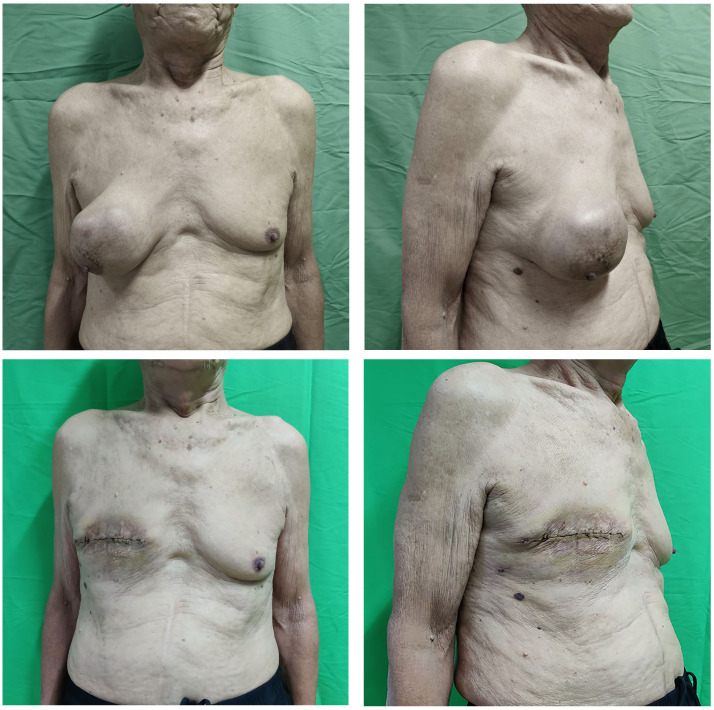
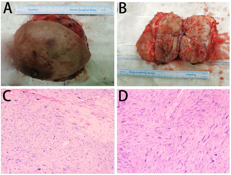
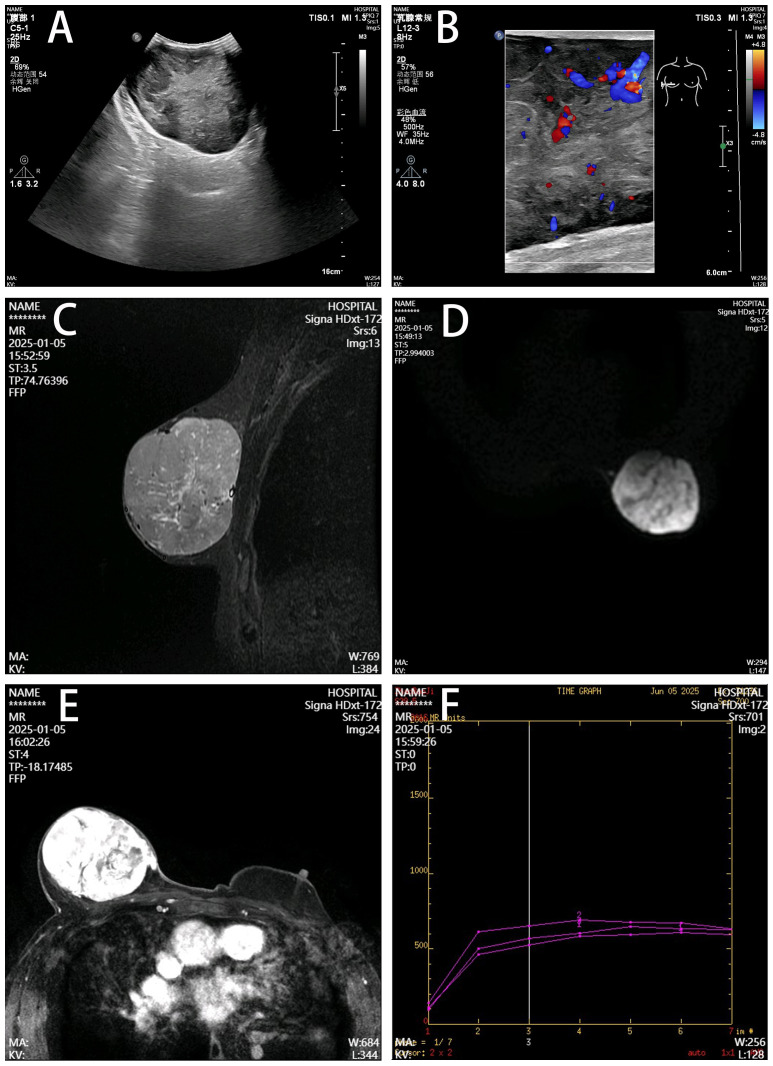
Primary breast leiomyosarcoma is an extremely rare malignancy originating from mesenchymal tissue. Fewer than 10 male cases have been reported globally. This paper reports an 84-year-old male patient. This represents the oldest reported case in the current literature. A painless, slowly enlarging mass was present in his right breast. The mass had a 10-year history. This contrasts sharply with the typically rapidly progressive pattern documented in previous literature. Clinical examination revealed a mobile mass measuring 12 cm × 10 cm in the right breast. No lymphadenopathy was detected. Ultrasound showed a hypoechoic lesion classified as BI-RADS 4a. Magnetic resonance imaging demonstrated plateau-type enhancement. The patient underwent simple mastectomy. Axillary lymph node dissection was not performed. Postoperative pathology and immunohistochemistry confirmed the diagnosis of breast leiomyosarcoma. The patient declined adjuvant radiotherapy. Follow-up at 6 months postoperatively showed no local recurrence or metastasis. This case indicates several points to clinicians. Immunohistochemistry serves as the cornerstone for diagnosing spindle cell tumors of the breast. R0 surgical resection constitutes the core approach for achieving cure. Decisions regarding adjuvant therapy require full consideration of host age and tumor biological behavior. The senescent microenvironment in elderly patients may suppress aggressive tumor progression.
期刊介绍:
Cancer Imaging and Diagnosis is dedicated to the publication of results from clinical and research studies applied to cancer diagnosis and treatment. The section aims to publish studies from the entire field of cancer imaging: results from routine use of clinical imaging in both radiology and nuclear medicine, results from clinical trials, experimental molecular imaging in humans and small animals, research on new contrast agents in CT, MRI, ultrasound, publication of new technical applications and processing algorithms to improve the standardization of quantitative imaging and image guided interventions for the diagnosis and treatment of cancer.

 求助内容:
求助内容: 应助结果提醒方式:


