Roberto Vega , Arun Nagdev , Masood Dehghan , Seyed Ehsan Seyed Bolouri , Brian Buchanan , Jeevesh Kapur , Jacob L. Jaremko , Dornoosh Zonoobi
{"title":"A wall tracking method to estimate ejection fraction from the parasternal long axis view in point of care ultrasound","authors":"Roberto Vega , Arun Nagdev , Masood Dehghan , Seyed Ehsan Seyed Bolouri , Brian Buchanan , Jeevesh Kapur , Jacob L. Jaremko , Dornoosh Zonoobi","doi":"10.1016/j.wfumbo.2025.100097","DOIUrl":null,"url":null,"abstract":"<div><h3>Objective:</h3><div>The left ventricular ejection fraction is a key metric for evaluating the systolic function in critically ill patients. Traditionally, it is computed using apical 2- and 4-chamber views in an echocardiogram; however, obtaining these views in an acutely ill patient in the emergency department is often difficult. A parasternal long-axis view, acquired with point of care ultrasound, is a faster and easier alternative. Unfortunately, the methods for estimating the ejection fraction from this view underperform when the left ventricular wall movement is not uniform or when its shape is not properly modeled as an ellipsoid. We propose a novel method that tracks the movement of the visible portions of the walls during the full cardiac cycle, and then estimates the ejection fraction based on that movement.</div></div><div><h3>Methods:</h3><div>We compared the performance of this method with the ejection fraction from the cardiology report on a dataset of 613 patients.</div></div><div><h3>Results:</h3><div>Our experiments showed an accuracy of 85% for identifying critically low values for ejection fraction (EF <span><math><mo><</mo></math></span> 30%) and 87% for abnormal ones (EF <span><math><mo><</mo></math></span> 50%). These values are comparable with the results obtained from the apical views and superior to current methods for the parasternal long-axis view.</div></div><div><h3>Conclusion:</h3><div>Since our method is fully automated, we expect that it can be adopted at scale in real-world clinical scenarios, giving practitioners a new tool to properly estimate the ejection fraction in clinically challenging scenarios.</div></div>","PeriodicalId":101281,"journal":{"name":"WFUMB Ultrasound Open","volume":"3 2","pages":"Article 100097"},"PeriodicalIF":0.0000,"publicationDate":"2025-10-10","publicationTypes":"Journal Article","fieldsOfStudy":null,"isOpenAccess":false,"openAccessPdf":"","citationCount":"0","resultStr":null,"platform":"Semanticscholar","paperid":null,"PeriodicalName":"WFUMB Ultrasound Open","FirstCategoryId":"1085","ListUrlMain":"https://www.sciencedirect.com/science/article/pii/S2949668325000199","RegionNum":0,"RegionCategory":null,"ArticlePicture":[],"TitleCN":null,"AbstractTextCN":null,"PMCID":null,"EPubDate":"","PubModel":"","JCR":"","JCRName":"","Score":null,"Total":0}
引用次数: 0
Abstract
Objective:
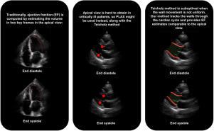
The left ventricular ejection fraction is a key metric for evaluating the systolic function in critically ill patients. Traditionally, it is computed using apical 2- and 4-chamber views in an echocardiogram; however, obtaining these views in an acutely ill patient in the emergency department is often difficult. A parasternal long-axis view, acquired with point of care ultrasound, is a faster and easier alternative. Unfortunately, the methods for estimating the ejection fraction from this view underperform when the left ventricular wall movement is not uniform or when its shape is not properly modeled as an ellipsoid. We propose a novel method that tracks the movement of the visible portions of the walls during the full cardiac cycle, and then estimates the ejection fraction based on that movement.
Methods:
We compared the performance of this method with the ejection fraction from the cardiology report on a dataset of 613 patients.
Results:
Our experiments showed an accuracy of 85% for identifying critically low values for ejection fraction (EF 30%) and 87% for abnormal ones (EF 50%). These values are comparable with the results obtained from the apical views and superior to current methods for the parasternal long-axis view.
Conclusion:
Since our method is fully automated, we expect that it can be adopted at scale in real-world clinical scenarios, giving practitioners a new tool to properly estimate the ejection fraction in clinically challenging scenarios.

 求助内容:
求助内容: 应助结果提醒方式:


