Effects of different reperfusion durations on neuronal mitochondrial ultrastructure following cerebral ischemia in diabetic rats: a transmission electron microscopy study.
{"title":"Effects of different reperfusion durations on neuronal mitochondrial ultrastructure following cerebral ischemia in diabetic rats: a transmission electron microscopy study.","authors":"Dan Wang, Gui-Sheng Chen","doi":"10.1186/s40001-025-03273-0","DOIUrl":null,"url":null,"abstract":"<p><strong>Objective: </strong>This study employed transmission electron microscopy (TEM) to characterize the temporal dynamics of neuronal mitochondrial ultrastructural alterations following cerebral ischemia-reperfusion (I/R) in diabetic rats.</p><p><strong>Methods: </strong>Male Wistar rats were divided into four groups: hyperglycemic, normoglycemic, hyperglycemic cerebral ischemia, and normoglycemic cerebral ischemia. Diabetic rat models were established via streptozotocin (STZ) induction. Cerebral ischemia was induced by bilateral common carotid artery occlusion combined with hypotension for 10 min, followed by reperfusion for 5 h, 1 day, or 7 days. Neurological deficits were evaluated using the Longa scoring criteria, and mitochondrial damage in the cortical region was assessed by TEM. Transmission electron micrographs were analyzed using ImageJ software (National Institutes of Health, USA).</p><p><strong>Results: </strong>(1) Diabetic rats exhibited exacerbated neuronal and mitochondrial damage compared with normoglycemic controls after I/R, with the most severe injury observed at 5 h of reperfusion. (2) Quantitative analysis revealed significantly greater mitochondrial swelling and cristae disruption in the hyperglycemic I/R group at all time points (p < 0.01). (3) Although prolonged reperfusion time correlated with gradual recovery of mitochondrial integrity, this recovery was significantly delayed and incomplete in diabetic rats compared with normoglycemic controls.</p><p><strong>Conclusions: </strong>(1) A diabetic cerebral I/R injury model was successfully established using STZ combined with bilateral carotid artery occlusion and hypotension. (2) Diabetes markedly exacerbates and prolongs mitochondrial damage following cerebral I/R. Impaired recovery of mitochondrial ultrastructure represents a critical determinant of stroke prognosis in diabetic patients, providing a well-defined therapeutic target for subsequent fundamental research and intervention strategies.</p>","PeriodicalId":11949,"journal":{"name":"European Journal of Medical Research","volume":"30 1","pages":"979"},"PeriodicalIF":3.4000,"publicationDate":"2025-10-15","publicationTypes":"Journal Article","fieldsOfStudy":null,"isOpenAccess":false,"openAccessPdf":"https://www.ncbi.nlm.nih.gov/pmc/articles/PMC12522883/pdf/","citationCount":"0","resultStr":null,"platform":"Semanticscholar","paperid":null,"PeriodicalName":"European Journal of Medical Research","FirstCategoryId":"3","ListUrlMain":"https://doi.org/10.1186/s40001-025-03273-0","RegionNum":3,"RegionCategory":"医学","ArticlePicture":[],"TitleCN":null,"AbstractTextCN":null,"PMCID":null,"EPubDate":"","PubModel":"","JCR":"Q2","JCRName":"MEDICINE, RESEARCH & EXPERIMENTAL","Score":null,"Total":0}
引用次数: 0
Abstract
Objective: This study employed transmission electron microscopy (TEM) to characterize the temporal dynamics of neuronal mitochondrial ultrastructural alterations following cerebral ischemia-reperfusion (I/R) in diabetic rats.
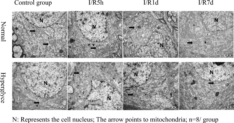
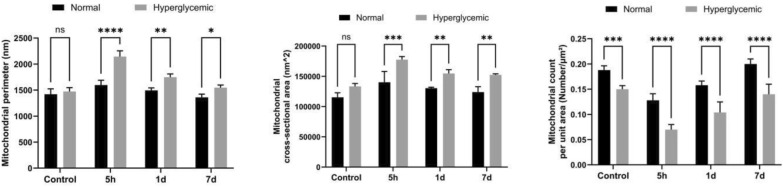
Methods: Male Wistar rats were divided into four groups: hyperglycemic, normoglycemic, hyperglycemic cerebral ischemia, and normoglycemic cerebral ischemia. Diabetic rat models were established via streptozotocin (STZ) induction. Cerebral ischemia was induced by bilateral common carotid artery occlusion combined with hypotension for 10 min, followed by reperfusion for 5 h, 1 day, or 7 days. Neurological deficits were evaluated using the Longa scoring criteria, and mitochondrial damage in the cortical region was assessed by TEM. Transmission electron micrographs were analyzed using ImageJ software (National Institutes of Health, USA).
Results: (1) Diabetic rats exhibited exacerbated neuronal and mitochondrial damage compared with normoglycemic controls after I/R, with the most severe injury observed at 5 h of reperfusion. (2) Quantitative analysis revealed significantly greater mitochondrial swelling and cristae disruption in the hyperglycemic I/R group at all time points (p < 0.01). (3) Although prolonged reperfusion time correlated with gradual recovery of mitochondrial integrity, this recovery was significantly delayed and incomplete in diabetic rats compared with normoglycemic controls.
Conclusions: (1) A diabetic cerebral I/R injury model was successfully established using STZ combined with bilateral carotid artery occlusion and hypotension. (2) Diabetes markedly exacerbates and prolongs mitochondrial damage following cerebral I/R. Impaired recovery of mitochondrial ultrastructure represents a critical determinant of stroke prognosis in diabetic patients, providing a well-defined therapeutic target for subsequent fundamental research and intervention strategies.
期刊介绍:
European Journal of Medical Research publishes translational and clinical research of international interest across all medical disciplines, enabling clinicians and other researchers to learn about developments and innovations within these disciplines and across the boundaries between disciplines. The journal publishes high quality research and reviews and aims to ensure that the results of all well-conducted research are published, regardless of their outcome.
 求助内容:
求助内容: 应助结果提醒方式:


