Demographic, Morphological, and Histopathological Characteristics of Melanoma and Nevi: Insights from Statistical Analysis and Machine Learning Models.
Blagjica Lazarova, Gordana Petrushevska, Zdenka Stojanovska, Stephen C Mullins
{"title":"Demographic, Morphological, and Histopathological Characteristics of Melanoma and Nevi: Insights from Statistical Analysis and Machine Learning Models.","authors":"Blagjica Lazarova, Gordana Petrushevska, Zdenka Stojanovska, Stephen C Mullins","doi":"10.3390/diagnostics15192499","DOIUrl":null,"url":null,"abstract":"<p><p><b>Background:</b> Early and accurate differentiation between melanomas and benign nevi is essential for making proper clinical decisions. This study aimed to identify clinical, morphological, and histopathological variables most strongly associated with melanoma, using both statistical and machine learning approaches. <b>Methods</b>: This study evaluated 184 melanocytic lesions using clinical, morphological, and histopathological parameters. Univariable analyses were performed in XLStat statistical software, version 2014.5.03, while multivariable machine learning models were developed in Jamovi (version 2.4). Five supervised algorithms (random forest, partial least squares, elastic net regression, conditional inference trees, and k-nearest neighbors) were compared using repeated cross-validation, with performance evaluated by accuracy, Kappa, sensitivity, specificity, F1 score, and calibration. <b>Results</b>: Univariable analysis identified significant differences between melanomas and nevi in age, horizontal diameter, gender, lesion location, and selected histopathological features (cytological and extracellular matrix changes, epidermal interactions). However, several associations weakened in multivariable analysis due to collinearity and overlapping effects. Using glmnet, the most influential independent predictors were cytological changes, horizontal diameter, epidermal interactions, and extracellular matrix features, alongside age, gender, and lesion location. The model achieved high discrimination (AUC = 0.97, 95% CI: 0.93-0.99) and accuracy (training: 95.3%; test: 92.6%), confirming robustness. <b>Conclusions</b>: Structured demographic, morphological, and histopathological data-particularly age, lesion size, cytological and extracellular matrix changes, and epidermal interactions-can effectively support classification of melanocytic lesions. Machine learning approaches (the glmnet model in our study) provide a reliable framework to evaluate such predictors and offer practical diagnostic support in dermatopathology.</p>","PeriodicalId":11225,"journal":{"name":"Diagnostics","volume":"15 19","pages":""},"PeriodicalIF":3.3000,"publicationDate":"2025-10-01","publicationTypes":"Journal Article","fieldsOfStudy":null,"isOpenAccess":false,"openAccessPdf":"https://www.ncbi.nlm.nih.gov/pmc/articles/PMC12524229/pdf/","citationCount":"0","resultStr":null,"platform":"Semanticscholar","paperid":null,"PeriodicalName":"Diagnostics","FirstCategoryId":"3","ListUrlMain":"https://doi.org/10.3390/diagnostics15192499","RegionNum":3,"RegionCategory":"医学","ArticlePicture":[],"TitleCN":null,"AbstractTextCN":null,"PMCID":null,"EPubDate":"","PubModel":"","JCR":"Q1","JCRName":"MEDICINE, GENERAL & INTERNAL","Score":null,"Total":0}
引用次数: 0
Abstract
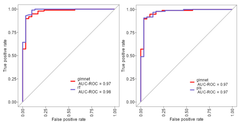
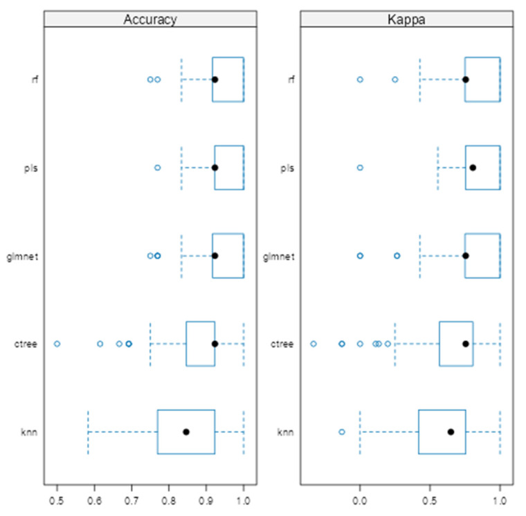
Background: Early and accurate differentiation between melanomas and benign nevi is essential for making proper clinical decisions. This study aimed to identify clinical, morphological, and histopathological variables most strongly associated with melanoma, using both statistical and machine learning approaches. Methods: This study evaluated 184 melanocytic lesions using clinical, morphological, and histopathological parameters. Univariable analyses were performed in XLStat statistical software, version 2014.5.03, while multivariable machine learning models were developed in Jamovi (version 2.4). Five supervised algorithms (random forest, partial least squares, elastic net regression, conditional inference trees, and k-nearest neighbors) were compared using repeated cross-validation, with performance evaluated by accuracy, Kappa, sensitivity, specificity, F1 score, and calibration. Results: Univariable analysis identified significant differences between melanomas and nevi in age, horizontal diameter, gender, lesion location, and selected histopathological features (cytological and extracellular matrix changes, epidermal interactions). However, several associations weakened in multivariable analysis due to collinearity and overlapping effects. Using glmnet, the most influential independent predictors were cytological changes, horizontal diameter, epidermal interactions, and extracellular matrix features, alongside age, gender, and lesion location. The model achieved high discrimination (AUC = 0.97, 95% CI: 0.93-0.99) and accuracy (training: 95.3%; test: 92.6%), confirming robustness. Conclusions: Structured demographic, morphological, and histopathological data-particularly age, lesion size, cytological and extracellular matrix changes, and epidermal interactions-can effectively support classification of melanocytic lesions. Machine learning approaches (the glmnet model in our study) provide a reliable framework to evaluate such predictors and offer practical diagnostic support in dermatopathology.
DiagnosticsBiochemistry, Genetics and Molecular Biology-Clinical Biochemistry
CiteScore
4.70
自引率
8.30%
发文量
2699
审稿时长
19.64 days
期刊介绍:
Diagnostics (ISSN 2075-4418) is an international scholarly open access journal on medical diagnostics. It publishes original research articles, reviews, communications and short notes on the research and development of medical diagnostics. There is no restriction on the length of the papers. Our aim is to encourage scientists to publish their experimental and theoretical research in as much detail as possible. Full experimental and/or methodological details must be provided for research articles.

 求助内容:
求助内容: 应助结果提醒方式:


