Keyi Zhang, Qi He, Yu Jin, Caihan Duan, Jun Liu, Chaoqun Han, Rong Lin
{"title":"Diagnostic Value of EUS-FNA in the Differential Diagnosis of Esophageal Strictures Lacking Typical Malignant Features.","authors":"Keyi Zhang, Qi He, Yu Jin, Caihan Duan, Jun Liu, Chaoqun Han, Rong Lin","doi":"10.3390/diagnostics15192470","DOIUrl":null,"url":null,"abstract":"<p><p><b>Background:</b> Esophageal strictures lacking typical malignant endoscopic features present a significant diagnostic challenge, often mimicking malignancy on imaging while concealing their true nature under regular white-light endoscopy. This study evaluated the utility of EUS-FNA in the differential diagnosis of such indeterminate strictures. <b>Methods:</b> We retrospectively analyzed 38 patients with suspicious malignant esophageal strictures indicated by CT but lacking definite malignant features on initial white-light gastroscopy. All patients underwent EUS-FNA for definitive pathological diagnosis. Clinicopathological data, imaging reports, endoscopic mucosal features, and procedural outcomes were assessed. <b>Results:</b> Among all 38 patients suspected of esophageal cancer by CT scan, 30 of them had malignant cytology results, including ESCC, EAC, metastatic cancer, and esophageal lymphoma. A total of 8 patients had benign findings, including esophageal tuberculosis, fungal esophagitis, eosinophilic esophagitis, and esophageal varices. Critically, EUS-FNA identified benign entities, such as eosinophilic esophagitis and esophageal tuberculosis masquerading as malignancy. CT features and mucosal features are also summarized and analyzed. <b>Conclusions:</b> EUS-FNA is a powerful tool for diagnosing esophageal strictures lacking typical malignant features. It reliably differentiates malignancy from challenging benign mimics, preventing misdiagnosis and guiding appropriate therapy. Clinicians should maintain a high suspicion for both occult malignancy and rare benign conditions in such stenotic lesions.</p>","PeriodicalId":11225,"journal":{"name":"Diagnostics","volume":"15 19","pages":""},"PeriodicalIF":3.3000,"publicationDate":"2025-09-26","publicationTypes":"Journal Article","fieldsOfStudy":null,"isOpenAccess":false,"openAccessPdf":"https://www.ncbi.nlm.nih.gov/pmc/articles/PMC12524196/pdf/","citationCount":"0","resultStr":null,"platform":"Semanticscholar","paperid":null,"PeriodicalName":"Diagnostics","FirstCategoryId":"3","ListUrlMain":"https://doi.org/10.3390/diagnostics15192470","RegionNum":3,"RegionCategory":"医学","ArticlePicture":[],"TitleCN":null,"AbstractTextCN":null,"PMCID":null,"EPubDate":"","PubModel":"","JCR":"Q1","JCRName":"MEDICINE, GENERAL & INTERNAL","Score":null,"Total":0}
引用次数: 0
Abstract
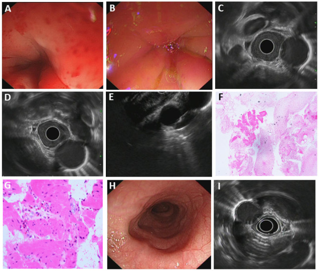
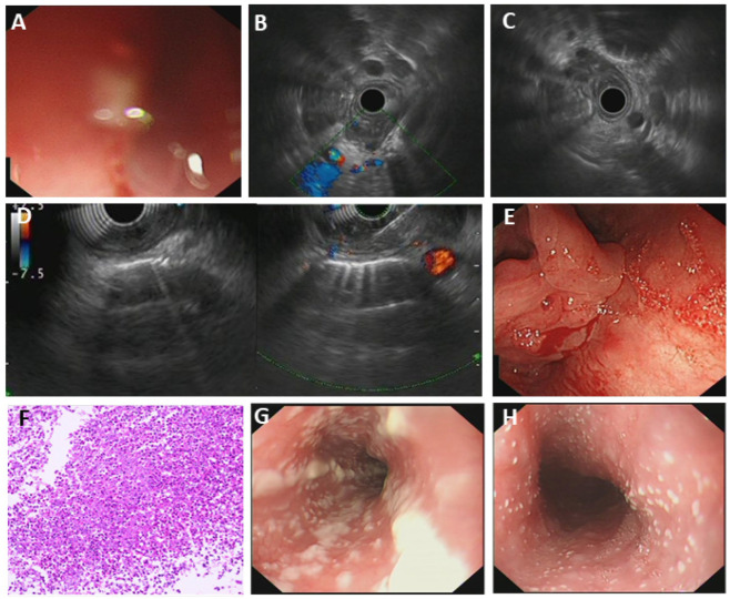
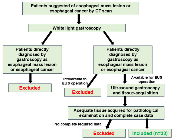
Background: Esophageal strictures lacking typical malignant endoscopic features present a significant diagnostic challenge, often mimicking malignancy on imaging while concealing their true nature under regular white-light endoscopy. This study evaluated the utility of EUS-FNA in the differential diagnosis of such indeterminate strictures. Methods: We retrospectively analyzed 38 patients with suspicious malignant esophageal strictures indicated by CT but lacking definite malignant features on initial white-light gastroscopy. All patients underwent EUS-FNA for definitive pathological diagnosis. Clinicopathological data, imaging reports, endoscopic mucosal features, and procedural outcomes were assessed. Results: Among all 38 patients suspected of esophageal cancer by CT scan, 30 of them had malignant cytology results, including ESCC, EAC, metastatic cancer, and esophageal lymphoma. A total of 8 patients had benign findings, including esophageal tuberculosis, fungal esophagitis, eosinophilic esophagitis, and esophageal varices. Critically, EUS-FNA identified benign entities, such as eosinophilic esophagitis and esophageal tuberculosis masquerading as malignancy. CT features and mucosal features are also summarized and analyzed. Conclusions: EUS-FNA is a powerful tool for diagnosing esophageal strictures lacking typical malignant features. It reliably differentiates malignancy from challenging benign mimics, preventing misdiagnosis and guiding appropriate therapy. Clinicians should maintain a high suspicion for both occult malignancy and rare benign conditions in such stenotic lesions.
DiagnosticsBiochemistry, Genetics and Molecular Biology-Clinical Biochemistry
CiteScore
4.70
自引率
8.30%
发文量
2699
审稿时长
19.64 days
期刊介绍:
Diagnostics (ISSN 2075-4418) is an international scholarly open access journal on medical diagnostics. It publishes original research articles, reviews, communications and short notes on the research and development of medical diagnostics. There is no restriction on the length of the papers. Our aim is to encourage scientists to publish their experimental and theoretical research in as much detail as possible. Full experimental and/or methodological details must be provided for research articles.

 求助内容:
求助内容: 应助结果提醒方式:


